19
views
0
recommends
+1 Recommend
0 collections
    0
    shares
      • Record: found
      • Abstract: found
      • Article: found
      Is Open Access

      The plasma membrane calcium ATPase 4 signalling in cardiac fibroblasts mediates cardiomyocyte hypertrophy

      research-article

      Read this article at

      Bookmark
          There is no author summary for this article yet. Authors can add summaries to their articles on ScienceOpen to make them more accessible to a non-specialist audience.

          Abstract

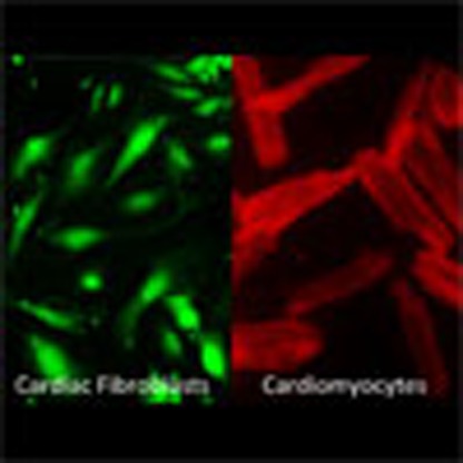
          The heart responds to pathological overload through myocyte hypertrophy. Here we show that this response is regulated by cardiac fibroblasts via a paracrine mechanism involving plasma membrane calcium ATPase 4 (PMCA4). Pmca4 deletion in mice, both systemically and specifically in fibroblasts, reduces the hypertrophic response to pressure overload; however, knocking out Pmca4 specifically in cardiomyocytes does not produce this effect. Mechanistically, cardiac fibroblasts lacking PMCA4 produce higher levels of secreted frizzled related protein 2 (sFRP2), which inhibits the hypertrophic response in neighbouring cardiomyocytes. Furthermore, we show that treatment with the PMCA4 inhibitor aurintricarboxylic acid (ATA) inhibits and reverses cardiac hypertrophy induced by pressure overload in mice. Our results reveal that PMCA4 regulates the development of cardiac hypertrophy and provide proof of principle for a therapeutic approach to treat this condition.

          Abstract

          Restricting hypertrophic heart growth in response to pathologic overload is an unmet therapeutic need. Here, the authors show that blocking Ca 2+ signaling controlled by the transport protein PMCA4 in cardiac fibroblasts enhances secretion of a potent Wnt signaling inhibitor, sFRP2, and prevents the development of pathologic cardiac hypertrophy in mice.

          Related collections

          Most cited references38

          • Record: found
          • Abstract: found
          • Article: not found

          In vivo reprogramming of murine cardiac fibroblasts into induced cardiomyocytes

          SUMMARY The reprogramming of adult cells into pluripotent cells or directly into alternative adult cell types holds great promise for regenerative medicine. We reported that cardiac fibroblasts, which represent 50% of the cells in the mammalian heart, can be directly reprogrammed to adult cardiomyocyte-like cells in vitro by the addition of Gata4, Mef2c and Tbx5 (GMT). Here, we use genetic lineage-tracing to show that resident non-myocytes in the murine heart can be reprogrammed into cardiomyocyte-like cells in vivo by local delivery of GMT after coronary ligation. Induced cardiomyocytes became bi-nucleate, assembled sarcomeres and had cardiomyocyte-like gene expression. Analysis of single cells revealed ventricular cardiomyocyte-like action potentials, beating upon electrical stimulation, and evidence of electrical coupling. In vivo delivery of GMT decreased infarct size and modestly attenuated cardiac dysfunction up to 3 months after coronary ligation. Delivery of the pro-angiogenic and fibroblast activating peptide, Thymosin β4, along with GMT, resulted in further improvements in scar area and cardiac function. These findings demonstrate that cardiac fibroblasts can be reprogrammed into cardiomyocyte-like cells in their native environment for potential regenerative purposes.
            Bookmark
            • Record: found
            • Abstract: not found
            • Article: not found

            Cardiac plasticity.

              Bookmark
              • Record: found
              • Abstract: found
              • Article: not found

              IL-33 and ST2 comprise a critical biomechanically induced and cardioprotective signaling system.

              ST2 is an IL-1 receptor family member with transmembrane (ST2L) and soluble (sST2) isoforms. sST2 is a mechanically induced cardiomyocyte protein, and serum sST2 levels predict outcome in patients with acute myocardial infarction or chronic heart failure. Recently, IL-33 was identified as a functional ligand of ST2L, allowing exploration of the role of ST2 in myocardium. We found that IL-33 was a biomechanically induced protein predominantly synthesized by cardiac fibroblasts. IL-33 markedly antagonized angiotensin II- and phenylephrine-induced cardiomyocyte hypertrophy. Although IL-33 activated NF-kappaB, it inhibited angiotensin II- and phenylephrine-induced phosphorylation of inhibitor of NF-kappa B alpha (I kappa B alpha) and NF-kappaB nuclear binding activity. sST2 blocked antihypertrophic effects of IL-33, indicating that sST2 functions in myocardium as a soluble decoy receptor. Following pressure overload by transverse aortic constriction (TAC), ST2(-/-) mice had more left ventricular hypertrophy, more chamber dilation, reduced fractional shortening, more fibrosis, and impaired survival compared with WT littermates. Furthermore, recombinant IL-33 treatment reduced hypertrophy and fibrosis and improved survival after TAC in WT mice, but not in ST2(-/-) littermates. Thus, IL-33/ST2 signaling is a mechanically activated, cardioprotective fibroblast-cardiomyocyte paracrine system, which we believe to be novel. IL-33 may have therapeutic potential for beneficially regulating the myocardial response to overload.
                Bookmark

                Author and article information

                Journal
                Nat Commun
                Nat Commun
                Nature Communications
                Nature Publishing Group
                2041-1723
                29 March 2016
                2016
                : 7
                : 11074
                Affiliations
                [1 ]Institute of Cardiovascular Sciences, University of Manchester , AV Hill Building, Manchester M13 9PT, UK
                [2 ]J David Gladstone Research Institutes , San Francisco, California 94158, USA
                [3 ]Faculty of Pharmacy, Zagazig University , Zagazig 44519, Egypt
                [4 ]Faculty of Life Sciences, University of Manchester , Manchester M13 9PT, UK
                Author notes
                [*]

                These authors contributed equally to this work.

                [†]

                These authors jointly supervised this work.

                Author information
                http://orcid.org/0000-0002-8435-808X
                Article
                ncomms11074
                10.1038/ncomms11074
                4820544
                27020607
                b82cd81a-8ec9-45e3-87cf-83e28162dd55
                Copyright © 2016, Nature Publishing Group, a division of Macmillan Publishers Limited. All Rights Reserved.

                This work is licensed under a Creative Commons Attribution 4.0 International License. The images or other third party material in this article are included in the article's Creative Commons license, unless indicated otherwise in the credit line; if the material is not included under the Creative Commons license, users will need to obtain permission from the license holder to reproduce the material. To view a copy of this license, visit http://creativecommons.org/licenses/by/4.0/

                History
                : 29 September 2015
                : 17 February 2016
                Categories
                Article

                Uncategorized
                Uncategorized

                Comments

                Comment on this article