21
views
0
recommends
+1 Recommend
0 collections
    0
    shares
      • Record: found
      • Abstract: found
      • Article: found
      Is Open Access

      Cellular Stresses and Stress Responses in the Pathogenesis of Insulin Resistance

      review-article
      Oxidative Medicine and Cellular Longevity
      Hindawi

      Read this article at

      Bookmark
          There is no author summary for this article yet. Authors can add summaries to their articles on ScienceOpen to make them more accessible to a non-specialist audience.

          Abstract

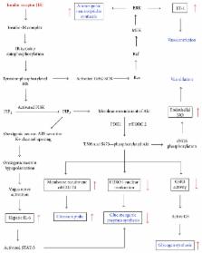
          Insulin resistance (IR), a key component of the metabolic syndrome, precedes the development of diabetes, cardiovascular disease, and Alzheimer's disease. Its etiological pathways are not well defined, although many contributory mechanisms have been established. This article summarizes such mechanisms into the hypothesis that factors like nutrient overload, physical inactivity, hypoxia, psychological stress, and environmental pollutants induce a network of cellular stresses, stress responses, and stress response dysregulations that jointly inhibit insulin signaling in insulin target cells including endothelial cells, hepatocytes, myocytes, hypothalamic neurons, and adipocytes. The insulin resistance-inducing cellular stresses include oxidative, nitrosative, carbonyl/electrophilic, genotoxic, and endoplasmic reticulum stresses; the stress responses include the ubiquitin-proteasome pathway, the DNA damage response, the unfolded protein response, apoptosis, inflammasome activation, and pyroptosis, while the dysregulated responses include the heat shock response, autophagy, and nuclear factor erythroid-2-related factor 2 signaling. Insulin target cells also produce metabolites that exacerbate cellular stress generation both locally and systemically, partly through recruitment and activation of myeloid cells which sustain a state of chronic inflammation. Thus, insulin resistance may be prevented or attenuated by multiple approaches targeting the different cellular stresses and stress responses.

          Related collections

          Most cited references321

          • Record: found
          • Abstract: found
          • Article: not found

          ER stress-induced cell death mechanisms.

          The endoplasmic-reticulum (ER) stress response constitutes a cellular process that is triggered by a variety of conditions that disturb folding of proteins in the ER. Eukaryotic cells have developed an evolutionarily conserved adaptive mechanism, the unfolded protein response (UPR), which aims to clear unfolded proteins and restore ER homeostasis. In cases where ER stress cannot be reversed, cellular functions deteriorate, often leading to cell death. Accumulating evidence implicates ER stress-induced cellular dysfunction and cell death as major contributors to many diseases, making modulators of ER stress pathways potentially attractive targets for therapeutics discovery. Here, we summarize recent advances in understanding the diversity of molecular mechanisms that govern ER stress signaling in health and disease. This article is part of a Special Section entitled: Cell Death Pathways. © 2013.
            Bookmark
            • Record: found
            • Abstract: found
            • Article: not found

            NLRP3 inflammasome activation results in hepatocyte pyroptosis, liver inflammation, and fibrosis in mice.

            Inflammasome activation plays a central role in the development of drug-induced and obesity-associated liver disease. However, the sources and mechanisms of inflammasome-mediated liver damage remain poorly understood. Our aim was to investigate the effect of NLRP3 inflammasome activation on the liver using novel mouse models. We generated global and myeloid cell-specific conditional mutant Nlrp3 knock-in mice expressing the D301N Nlrp3 mutation (ortholog of D303N in human NLRP3), resulting in a hyperactive NLRP3. To study the presence and significance of NLRP3-initiated pyroptotic cell death, we separated hepatocytes from nonparenchymal cells and developed a novel flow-cytometry-based (fluorescence-activated cell sorting; FACS) strategy to detect and quantify pyroptosis in vivo based on detection of active caspase 1 (Casp1)- and propidium iodide (PI)-positive cells. Liver inflammation was quantified histologically by FACS and gene expression analysis. Liver fibrosis was assessed by Sirius Red staining and quantitative polymerase chain reaction for markers of hepatic stellate cell (HSC) activation. NLRP3 activation resulted in shortened survival, poor growth, and severe liver inflammation; characterized by neutrophilic infiltration and HSC activation with collagen deposition in the liver. These changes were partially attenuated by treatment with anakinra, an interleukin-1 receptor antagonist. Notably, hepatocytes from global Nlrp3-mutant mice showed marked hepatocyte pyroptotic cell death, with more than a 5-fold increase in active Casp1/PI double-positive cells. Myeloid cell-restricted mutant NLRP3 activation resulted in a less-severe liver phenotype in the absence of detectable pyroptotic hepatocyte cell death. Our data demonstrate that global and, to a lesser extent, myeloid-specific NLRP3 inflammasome activation results in severe liver inflammation and fibrosis while identifying hepatocyte pyroptotic cell death as a novel mechanism of NLRP3-mediated liver damage. © 2014 by the American Association for the Study of Liver Diseases.
              Bookmark
              • Record: found
              • Abstract: found
              • Article: not found

              A crucial role for adipose tissue p53 in the regulation of insulin resistance.

              Various stimuli, such as telomere dysfunction and oxidative stress, can induce irreversible cell growth arrest, which is termed 'cellular senescence'. This response is controlled by tumor suppressor proteins such as p53 and pRb. There is also evidence that senescent cells promote changes related to aging or age-related diseases. Here we show that p53 expression in adipose tissue is crucially involved in the development of insulin resistance, which underlies age-related cardiovascular and metabolic disorders. We found that excessive calorie intake led to the accumulation of oxidative stress in the adipose tissue of mice with type 2 diabetes-like disease and promoted senescence-like changes, such as increased activity of senescence-associated beta-galactosidase, increased expression of p53 and increased production of proinflammatory cytokines. Inhibition of p53 activity in adipose tissue markedly ameliorated these senescence-like changes, decreased the expression of proinflammatory cytokines and improved insulin resistance in mice with type 2 diabetes-like disease. Conversely, upregulation of p53 in adipose tissue caused an inflammatory response that led to insulin resistance. Adipose tissue from individuals with diabetes also showed senescence-like features. Our results show a previously unappreciated role of adipose tissue p53 expression in the regulation of insulin resistance and suggest that cellular aging signals in adipose tissue could be a new target for the treatment of diabetes (pages 996-967).
                Bookmark

                Author and article information

                Contributors
                Journal
                Oxid Med Cell Longev
                Oxid Med Cell Longev
                OMCL
                Oxidative Medicine and Cellular Longevity
                Hindawi
                1942-0900
                1942-0994
                2018
                9 July 2018
                : 2018
                : 4321714
                Affiliations
                Department of Food Science and Technology, Jomo Kenyatta University of Agriculture and Technology, P.O. Box 62000, Nairobi 00200, Kenya
                Author notes

                Academic Editor: Claudio Cabello-Verrugio

                Author information
                http://orcid.org/0000-0003-2078-6496
                Article
                10.1155/2018/4321714
                6079365
                30116482
                01593125-d958-40c3-b578-736278d13d16
                Copyright © 2018 Arnold N. Onyango.

                This is an open access article distributed under the Creative Commons Attribution License, which permits unrestricted use, distribution, and reproduction in any medium, provided the original work is properly cited.

                History
                : 1 January 2018
                : 18 February 2018
                Categories
                Review Article

                Molecular medicine
                Molecular medicine

                Comments

                Comment on this article