11
views
0
recommends
+1 Recommend
0 collections
    0
    shares
      • Record: found
      • Abstract: found
      • Article: found
      Is Open Access

      Comparison of echocardiographic indices of right ventricular systolic function and ejection fraction obtained with continuous thermodilution in critically ill patients

      research-article

      Read this article at

      Bookmark
          There is no author summary for this article yet. Authors can add summaries to their articles on ScienceOpen to make them more accessible to a non-specialist audience.

          Abstract

          Background

          Though echocardiographic evaluation assesses the right ventricular systolic function, which of the existing parameters best reflects the right ventricular ejection fraction (RVEF) in the critically ill patients is still uncertain. We aimed to determine the relationship between echocardiographic indices of right ventricular systolic function and RVEF.

          Methods

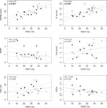
          Prospective observational study was conducted in a mixed Surgical Intensive Care Unit (Hôpital Lariboisière, Paris, France) from November 2017 to November 2018. All critically ill patients monitored with a pulmonary artery catheter were assessed. We collected echocardiographic indices of right ventricular function (tricuspid annular plane systolic excursion, TAPSE; peak systolic velocity of pulsed tissue Doppler at lateral tricuspid annulus, S′; fractional area change, FAC; right ventricular index of myocardial performance, RIMP; isovolumic acceleration, IVA; end-diastolic diameter ratio, EDDr) and compared them with the RVEF obtained from continuous volumetric pulmonary artery catheter.

          Results

          Twenty-five patients were analyzed. Admission diagnosis was acute heart failure in 11 patients and septic shock in 14 patients. Median age was 70 years [57–80], norepinephrine median dose was 0.29 μg/kg/min [0.14–0.50], median Sequential Organ Failure Assessment score was 12 [10–14], and mortality at day 28 was 56%. When compared to RVEF, TAPSE had the highest correlation coefficient (rho = 0.78, 95% CI 0.52 to 0.89, p < 0.001). S′ was also correlated to RVEF (rho = 0.64, 95% CI 0.60 to 0.80, p = 0.001) whereas FAC, RIMP, IVA, and EDDr did not. TAPSE lower than 16 mm, S′ lower than 11 cm/s, and EDDr higher than 1 were always associated with a reduced RVEF.

          Conclusions

          We found that amongst indices of right ventricular systolic function, TAPSE and S′ were well correlated with thermodilution-derived RVEF in critically ill patients.

          Electronic supplementary material

          The online version of this article (10.1186/s13054-019-2582-7) contains supplementary material, which is available to authorized users.

          Related collections

          Most cited references37

          • Record: found
          • Abstract: found
          • Article: not found

          Evaluation and Management of Right-Sided Heart Failure: A Scientific Statement From the American Heart Association

          The diverse causes of right-sided heart failure (RHF) include, among others, primary cardiomyopathies with right ventricular (RV) involvement, RV ischemia and infarction, volume loading caused by cardiac lesions associated with congenital heart disease and valvular pathologies, and pressure loading resulting from pulmonic stenosis or pulmonary hypertension from a variety of causes, including left-sided heart disease. Progressive RV dysfunction in these disease states is associated with increased morbidity and mortality. The purpose of this scientific statement is to provide guidance on the assessment and management of RHF.
            Bookmark
            • Record: found
            • Abstract: found
            • Article: not found

            Contemporary management of acute right ventricular failure: a statement from the Heart Failure Association and the Working Group on Pulmonary Circulation and Right Ventricular Function of the European Society of Cardiology.

            Acute right ventricular (RV) failure is a complex clinical syndrome that results from many causes. Research efforts have disproportionately focused on the failing left ventricle, but recently the need has been recognized to achieve a more comprehensive understanding of RV anatomy, physiology, and pathophysiology, and of management approaches. Right ventricular mechanics and function are altered in the setting of either pressure overload or volume overload. Failure may also result from a primary reduction of myocardial contractility owing to ischaemia, cardiomyopathy, or arrhythmia. Dysfunction leads to impaired RV filling and increased right atrial pressures. As dysfunction progresses to overt RV failure, the RV chamber becomes more spherical and tricuspid regurgitation is aggravated, a cascade leading to increasing venous congestion. Ventricular interdependence results in impaired left ventricular filling, a decrease in left ventricular stroke volume, and ultimately low cardiac output and cardiogenic shock. Identification and treatment of the underlying cause of RV failure, such as acute pulmonary embolism, acute respiratory distress syndrome, acute decompensation of chronic pulmonary hypertension, RV infarction, or arrhythmia, is the primary management strategy. Judicious fluid management, use of inotropes and vasopressors, assist devices, and a strategy focusing on RV protection for mechanical ventilation if required all play a role in the clinical care of these patients. Future research should aim to address the remaining areas of uncertainty which result from the complexity of RV haemodynamics and lack of conclusive evidence regarding RV-specific treatment approaches.
              Bookmark
              • Record: found
              • Abstract: found
              • Article: not found

              Traditional and innovative echocardiographic parameters for the analysis of right ventricular performance in comparison with cardiac magnetic resonance.

              Right ventricle fractional area change (RVFAC), tissue Doppler and M-mode measurements of tricuspid systolic motion [tricuspid Sm and tricuspid annular plane systolic excursion (TAPSE)], and 3D echocardiography are the current non-invasive methods for the quantification of RV systolic function; RV deformation analysis by speckle-tracking echocardiography (STE) has recently allowed the analysis of RV performance. Using cardiac magnetic resonance (CMR) as the reference standard, this study aimed at exploring the correlation between the traditional (fractional shortening, s'RV, TAPSE) and innovative (strain) echocardiographic parameters and RV ejection fraction (RVEF) measured by CMR.
                Bookmark

                Author and article information

                Contributors
                romain.barthelemy@aphp.fr
                royxavier75@gmail.com
                tsjava@outlook.com
                alexandre.mebazaa@aphp.fr
                benjamin.chousterman@aphp.fr
                Journal
                Crit Care
                Critical Care
                BioMed Central (London )
                1364-8535
                1466-609X
                13 September 2019
                13 September 2019
                2019
                : 23
                : 312
                Affiliations
                [1 ]ISNI 0000 0000 9725 279X, GRID grid.411296.9, Department of Anaesthesia and Critical Care, Lariboisière Hospital, DMU Parabol, APHP.Nord, ; Paris, France
                [2 ]ISNI 0000000121866389, GRID grid.7429.8, Inserm UMR-S942, Mascot, ; Paris, France
                [3 ]ISNI 0000 0001 2171 2558, GRID grid.5842.b, Université de Paris, ; Paris, France
                [4 ]ISNI 0000 0000 9725 279X, GRID grid.411296.9, Réanimation Chirurgical Polyvalente, Hôpital Lariboisière, ; 2 rue Ambroise Paré, 75475 Paris Cedex 10, France
                Author information
                http://orcid.org/0000-0002-6493-4326
                Article
                2582
                10.1186/s13054-019-2582-7
                6743193
                31519203
                017e5962-7aa0-4ad3-be05-dbf2c818a685
                © The Author(s). 2019

                Open AccessThis article is distributed under the terms of the Creative Commons Attribution 4.0 International License ( http://creativecommons.org/licenses/by/4.0/), which permits unrestricted use, distribution, and reproduction in any medium, provided you give appropriate credit to the original author(s) and the source, provide a link to the Creative Commons license, and indicate if changes were made. The Creative Commons Public Domain Dedication waiver ( http://creativecommons.org/publicdomain/zero/1.0/) applies to the data made available in this article, unless otherwise stated.

                History
                : 24 April 2019
                : 27 August 2019
                Categories
                Research
                Custom metadata
                © The Author(s) 2019

                Emergency medicine & Trauma
                critical illness,ventricular dysfunction,right,catheterization,swan-ganz,thermodilution,echocardiography,2d,doppler

                Comments

                Comment on this article