20
views
0
recommends
+1 Recommend
0 collections
    0
    shares
      • Record: found
      • Abstract: found
      • Article: found
      Is Open Access

      Choroidal thickness in patients with diabetic retinopathy

      research-article

      Read this article at

      Bookmark
          There is no author summary for this article yet. Authors can add summaries to their articles on ScienceOpen to make them more accessible to a non-specialist audience.

          Abstract

          Purpose

          The aim of the study reported here was to assess choroidal thickness (CT) and central macular thickness (CMT) in patients with diabetic retinopathy.

          Materials and methods

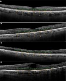
          A total of 151 eyes from 80 patients from the retina department of Istanbul Training and Research Hospital who had type 2 diabetes mellitus with diabetic retinopathy were studied retrospectively in this cross-sectional research. Patients were divided into three groups: mild–moderate nonproliferative diabetic retinopathy without macular edema (NPDR), mild–moderate nonproliferative diabetic retinopathy with macular edema (DME), and proliferative diabetic retinopathy (PDR). In addition, 40 eyes of 20 healthy individuals comprised a control group. Choroidal thickness was measured from the posterior edge of the retinal pigment epithelium to the choroid/sclera junction at 500-μm intervals up to 1,500 μm temporal and nasal to the fovea. The CMT measurement was obtained for each eye. Serum hemoglobin A 1c (HbA 1c) levels were measured.

          Results

          The study included 191 eyes, comprising 151 eyes of 80 patients and 40 eyes of 20 healthy individuals. Of the 151 patient eyes, 61 had NPDR, 62 had PDR, and 28 eyes had DME. There was no statistically significant difference in age between the groups ( P>0.05). In both the PDR and DME groups, the CT was statistically significantly decreased compared with the control group ( P<0.001, P<0.001 for the PDR and DME groups, respectively). The mean CMT in the DME group was increased significantly compared with both the NPDR and PDR groups ( P<0.001, P<0.001, respectively). In all three groups, serum HbA 1c levels were found to be increased significantly compared with the control group ( P=0.000). We found a statistically weak–moderate negative correlation between central macular and foveal CT ( r=−289, P=0.000). There was a statistically strong correlation between CMT and HbA 1c levels ( r=0.577, P=0.483) and a statistically weak–moderate negative correlation between the central CT and HbA 1c levels ( r=−0.331, P<0.001).

          Conclusion

          Diabetes changes the CT. CT was found to be significantly decreased in the DME and PDR groups.

          Related collections

          Most cited references11

          • Record: found
          • Abstract: found
          • Article: not found

          Choroidal thickness in normal eyes measured using Cirrus HD optical coherence tomography.

          To examine choroidal thickness and area in healthy eyes using spectral-domain optical coherence tomography (SD-OCT). Retrospective, observational case series. Thirty-four eyes (34 subjects), with no retinal or choroidal disease, underwent high-definition raster scanning using SD-OCT with frame enhancement software. Choroidal thickness was measured from the posterior edge of the retinal pigment epithelium to the choroid/sclera junction at 500-microm intervals up to 2500 microm temporal and nasal to the fovea. The central 1-mm area of the choroid was also measured, along with foveal thickness of the retina. All measurements were performed by 2 independent observers. Statistical analysis was used to correlate inter-observer findings, choroidal thickness and area measurements with age, and choroidal thickness with retinal foveal thickness. The 34 subjects had a mean age of 51.1 years. Reliable measurements of choroidal thickness were obtainable in 74% of eyes examined. Choroidal thickness and area measurements had strong inter-observer correlation (r = 0.92, P < .0001 and r = 0.93, P < .0001 respectively). Area had a moderate negative correlation with age (r = -0.62, P < .0001) that was comparable to the correlation between mean subfoveal choroidal thickness and age (r = -0.61, P < .0001). Retinal and choroidal thickness were found to be poorly correlated (r = -0.23, P = .18). Mean choroidal thickness showed a pattern of thinnest choroid nasally, thickening in the subfoveal region, and then thinning again temporally. Mean subfoveal choroidal thickness was found to be 272 microm (SD, +/- 81 microm). Choroidal thickness can be measured using SD-OCT high-definition raster scans in the majority of eyes. Choroidal thickness across the macula demonstrates a thin choroid nasally, thickest subfoveally, and again thinner temporally, and a trend toward decreasing choroidal thickness with age. Copyright (c) 2010 Elsevier Inc. All rights reserved.
            Bookmark
            • Record: found
            • Abstract: found
            • Article: not found

            Changes in choroidal thickness in relation to the severity of retinopathy and macular edema in type 2 diabetic patients.

            To assess changes in choroidal thickness in type 2 diabetic patients with diabetic retinopathy (DR) and diabetic macular edema (DME) using enhanced-depth imaging spectral domain optical coherence tomography (EDI-OCT).
              Bookmark
              • Record: found
              • Abstract: found
              • Article: not found

              Choroidal thickness in patients with diabetic retinopathy analyzed by spectral-domain optical coherence tomography.

              This study was designed to examine choroidal thickness in patients with diabetes using spectral-domain optical coherence tomography. Forty-nine patients (49 eyes) with diabetes and 24 age-matched normal subjects underwent high-definition raster scanning using spectral-domain optical coherence tomography with frame enhancement software. Patients with diabetes were classified into 3 groups: 11 patients with mild or moderate nonproliferative diabetic retinopathy and no macular edema, 18 patients with nonproliferative diabetic retinopathy and diabetic macular edema, and 20 patients with treated proliferative diabetic retinopathy and no diabetic macular edema (treated proliferative diabetic retinopathy). Choroidal thickness was measured from the posterior edge of the retinal pigment epithelium to the choroid/sclera junction at 500-μm intervals up to 2,500 μm temporal and nasal to the fovea. Reliable measurements of choroidal thickness were obtainable in 75.3% of eyes examined. Mean choroidal thickness showed a pattern of thinnest choroid nasally, thickening in the subfoveal region, and thinning again temporally in normal subjects and patients with diabetes. Mean subfoveal choroidal thickness was thinner in patients with diabetic macular edema (63.3 μm, 27.2%, P < 0.05) or treated proliferative diabetic retinopathy (69.6 μm, 30.0%, P < 0.01), compared with normal subjects. There was no difference between nonproliferative diabetic retinopathy and normal subjects. Choroidal thickness is altered in diabetes and may be related to the severity of retinopathy. Presence of diabetic macular edema is associated with a significant decrease in the choroidal thickness.
                Bookmark

                Author and article information

                Journal
                Clin Ophthalmol
                Clin Ophthalmol
                Clinical Ophthalmology
                Clinical Ophthalmology (Auckland, N.Z.)
                Dove Medical Press
                1177-5467
                1177-5483
                2014
                27 March 2014
                : 8
                : 637-642
                Affiliations
                Department of Ophthalmology, Istanbul Education and Research Hospital, Istanbul, Turkey
                Author notes
                Correspondence: Erkan Ünsal, Istanbul Training and Research Hospital, Kasap Ilyas Mah Org Abdurrahman Nafiz, Gurman Cd, PK 34098, Fatih, Istanbul, Turkey, Tel +90 505 238 2250, Fax +90 212 459 6145, Email erkanunsal@ 123456gmail.com
                Article
                opth-8-637
                10.2147/OPTH.S59395
                3971934
                24707168
                03fa48eb-b77e-4d03-bb45-1a3b68e83976
                © 2014 Ünsal et al. This work is published by Dove Medical Press Limited, and licensed under Creative Commons Attribution – Non Commercial (unported, v3.0) License

                The full terms of the License are available at http://creativecommons.org/licenses/by-nc/3.0/. Non-commercial uses of the work are permitted without any further permission from Dove Medical Press Limited, provided the work is properly attributed.

                History
                Categories
                Original Research

                Ophthalmology & Optometry
                choroidal thickness,diabetic retinopathy,optical coherence tomography

                Comments

                Comment on this article