14
views
0
recommends
+1 Recommend
0 collections
    0
    shares
      • Record: found
      • Abstract: found
      • Article: found
      Is Open Access

      Evidence for a Resting State Network Abnormality in Adults Who Stutter

      research-article

      Read this article at

      Bookmark
          There is no author summary for this article yet. Authors can add summaries to their articles on ScienceOpen to make them more accessible to a non-specialist audience.

          Abstract

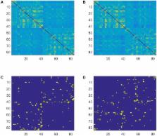
          Neural network-based investigations of stuttering have begun to provide a possible integrative account for the large number of brain-based anomalies associated with stuttering. Here we used resting-state EEG to investigate functional brain networks in adults who stutter ( AWS). Participants were 19 AWS and 52 age-, and gender-matched normally fluent speakers. EEGs were recorded and connectivity matrices were generated by LORETA in the theta (4–8 Hz), alpha (8–12 Hz), beta1 (12–20 Hz), and beta2 (20–30 Hz) bands. Small-world propensity ( SWP), shortest path, and clustering coefficients were computed for weighted graphs. Minimum spanning tree analysis was also performed and measures were compared by non-parametric permutation test. The results show that small-world topology was evident in the functional networks of all participants. Three graph indices (diameter, clustering coefficient, and shortest path) exhibited significant differences between groups in the theta band and one [maximum betweenness centrality ( BC)] measure was significantly different between groups in the beta2 band. AWS show higher BC than control in right temporal and inferior frontal areas and lower BC in the right primary motor cortex. Abnormal functional networks during rest state suggest an anomaly of DMN activity in AWS. Furthermore, functional segregation/integration deficits in the theta network are evident in AWS. These deficits reinforce the hypothesis that there is a neural basis for abnormal executive function in AWS. Increased beta2 BC in the right speech–motor related areas confirms previous evidence that right audio–speech areas are over-activated in AWS. Decreased beta2 BC in the right primary motor cortex is discussed in relation to abnormal neural mechanisms associated with time perception in AWS.

          Related collections

          Most cited references84

          • Record: found
          • Abstract: found
          • Article: not found

          Small-world brain networks.

          Many complex networks have a small-world topology characterized by dense local clustering or cliquishness of connections between neighboring nodes yet a short path length between any (distant) pair of nodes due to the existence of relatively few long-range connections. This is an attractive model for the organization of brain anatomical and functional networks because a small-world topology can support both segregated/specialized and distributed/integrated information processing. Moreover, small-world networks are economical, tending to minimize wiring costs while supporting high dynamical complexity. The authors introduce some of the key mathematical concepts in graph theory required for small-world analysis and review how these methods have been applied to quantification of cortical connectivity matrices derived from anatomical tract-tracing studies in the macaque monkey and the cat. The evolution of small-world networks is discussed in terms of a selection pressure to deliver cost-effective information-processing systems. The authors illustrate how these techniques and concepts are increasingly being applied to the analysis of human brain functional networks derived from electroencephalography/magnetoencephalography and fMRI experiments. Finally, the authors consider the relevance of small-world models for understanding the emergence of complex behaviors and the resilience of brain systems to pathological attack by disease or aberrant development. They conclude that small-world models provide a powerful and versatile approach to understanding the structure and function of human brain systems.
            Bookmark
            • Record: found
            • Abstract: found
            • Article: not found

            What makes us tick? Functional and neural mechanisms of interval timing.

            Time is a fundamental dimension of life. It is crucial for decisions about quantity, speed of movement and rate of return, as well as for motor control in walking, speech, playing or appreciating music, and participating in sports. Traditionally, the way in which time is perceived, represented and estimated has been explained using a pacemaker-accumulator model that is not only straightforward, but also surprisingly powerful in explaining behavioural and biological data. However, recent advances have challenged this traditional view. It is now proposed that the brain represents time in a distributed manner and tells the time by detecting the coincidental activation of different neural populations.
              Bookmark
              • Record: found
              • Abstract: found
              • Article: not found

              Low resolution electromagnetic tomography: a new method for localizing electrical activity in the brain.

              This paper presents a new method for localizing the electric activity in the brain based on multichannel surface EEG recordings. In contrast to the models presented up to now the new method does not assume a limited number of dipolar point sources nor a distribution on a given known surface, but directly computes a current distribution throughout the full brain volume. In order to find a unique solution for the 3-dimensional distribution among the infinite set of different possible solutions, the method assumes that neighboring neurons are simultaneously and synchronously activated. The basic assumption rests on evidence from single cell recordings in the brain that demonstrates strong synchronization of adjacent neurons. In view of this physiological consideration the computational task is to select the smoothest of all possible 3-dimensional current distributions, a task that is a common procedure in generalized signal processing. The result is a true 3-dimensional tomography with the characteristic that localization is preserved with a certain amount of dispersion, i.e., it has a relatively low spatial resolution. The new method, which we call Low Resolution Electromagnetic Tomography (LORETA) is illustrated with two different sets of evoked potential data, the first showing the tomography of the P100 component to checkerboard stimulation of the left, right, upper and lower hemiretina, and the second showing the results for the auditory N100 component and the two cognitive components CNV and P300. A direct comparison of the tomography results with those obtained from fitting one and two dipoles illustrates that the new method provides physiologically meaningful results while dipolar solutions fail in many situations. In the case of the cognitive components, the method offers new hypotheses on the location of higher cognitive functions in the brain.
                Bookmark

                Author and article information

                Contributors
                Journal
                Front Integr Neurosci
                Front Integr Neurosci
                Front. Integr. Neurosci.
                Frontiers in Integrative Neuroscience
                Frontiers Media S.A.
                1662-5145
                27 April 2018
                2018
                : 12
                : 16
                Affiliations
                [1] 1Cognitive Neuroscience Laboratory, University of Tabriz , Tabriz, Iran
                [2] 2Iranian Neuro-wave Laboratory, Center of Isfahan , Isfahan, Iran
                [3] 3Department of Physics, School of Basic Science, Babol Noshirvani University of Technology , Babol, Iran
                [4] 4Department of Cognitive Science, Faculty of Human Sciences, Macquarie University , Sydney, NSW, Australia
                [5] 5ARC Centre of Excellence in Cognition and its Disorders, Macquarie University , Sydney, NSW, Australia
                Author notes

                Edited by: James Danckert, University of Waterloo, Canada

                Reviewed by: Gennady Knyazev, State Scientific-Research Institute of Physiology & Basic Medicine, Russia; Irene Messina, Università degli Studi di Padova, Italy

                Article
                10.3389/fnint.2018.00016
                5934488
                049a4b56-7098-4f48-b71e-fa30ca4d49bd
                Copyright © 2018 Ghaderi, Andevari and Sowman.

                This is an open-access article distributed under the terms of the Creative Commons Attribution License (CC BY). The use, distribution or reproduction in other forums is permitted, provided the original author(s) and the copyright owner are credited and that the original publication in this journal is cited, in accordance with accepted academic practice. No use, distribution or reproduction is permitted which does not comply with these terms.

                History
                : 24 January 2018
                : 10 April 2018
                Page count
                Figures: 3, Tables: 0, Equations: 1, References: 102, Pages: 10, Words: 0
                Categories
                Neuroscience
                Original Research

                Neurosciences
                stuttering,functional brain networks,minimum spanning tree,executive function,time perception

                Comments

                Comment on this article