2
views
0
recommends
+1 Recommend
0 collections
    0
    shares
      • Record: found
      • Abstract: found
      • Article: found
      Is Open Access

      Pulmonary embolism as the initial manifestation of right atrial myxoma : A case report and review of the literature

      case-report
      , MD a , , MD b , , MD c , , MD a , , MD a , , MD a ,
      Medicine
      Wolters Kluwer Health
      atrial myxoma, diagnosis, pulmonary embolism, treatment

      Read this article at

      Bookmark
          There is no author summary for this article yet. Authors can add summaries to their articles on ScienceOpen to make them more accessible to a non-specialist audience.

          Abstract

          Rationale:

          Pulmonary embolisms (PEs) are caused by emboli, which mostly originate from deep venous thrombi that travel to and suddenly block the pulmonary arteries. The emboli are usually thrombi, and right atrial myxoma emboli are rare.

          Patient concerns:

          A 55-year-old man presented with shortness of breath and syncope. We proceeded with computed tomography pulmonary angiography (CTPA) and transthoracic echocardiogram (TTE), the results of which suggested that the diagnosis was a right atrial mass.

          Diagnosis:

          A definitive diagnosis compatible with a right atrial myxoma (RAM) with tumoral pulmonary emboli after surgical excision was made.

          Intervention:

          Right atrial and pulmonary artery embolectomy.

          Outcomes:

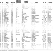
          The patient followed an uneventful course during the 6 years of follow-up after surgery. According to a review of the literature, RAMs are often not diagnosed in a timely manner or even go completely undiagnosed. TTE, transesophageal echocardiography (TEE), CT, magnetic resonance imaging (MRI), and positron emission tomography/computed tomography may be helpful in the preoperative diagnosis. Surgical removal of the masses from the atrium and pulmonary arteries was relatively uneventful.

          Lessons:

          RAMs should be considered unlikely reasons for fatal pulmonary embolisms.

          Related collections

          Most cited references61

          • Record: found
          • Abstract: found
          • Article: not found

          Cardiac myxomas.

          K Reynen (1995)
          Although cardiac myxomas are histologically benign, they may be lethal because of their strategic position. They can mimic not only every cardiac disease but also infective, immunologic, and malignant processes. Myxomas must therefore be included in the differential diagnosis of valvular heart disease, cardiac insufficiency, cardiomegaly, bacterial endocarditis, disturbances of ventricular and supraventricular rhythm, syncope, and systemic or pulmonary embolism. The symptoms depend on the size, mobility, and location of the tumor. Echocardiography, including the transesophageal approach, is the most important means of diagnosis; CT and MRI may also be helpful. Coronary arteriography in patients over 40 years of age is generally required to rule out concomitant coronary artery disease. Surgical removal of the tumor should be performed as soon as possible; the long-term prognosis is excellent, and recurrences are rare. In follow-up examinations as well, echocardiography is essential.
            Bookmark
            • Record: found
            • Abstract: found
            • Article: not found

            Clinical presentation of left atrial cardiac myxoma. A series of 112 consecutive cases.

            We analyzed a series of 112 consecutive cases of left atrial myxoma diagnosed in a single French hospital (72 women and 40 men; age range, 5-84 yr) over 40 years, from 1959 to 1998. Symptoms of mitral valve obstruction, the first arm of the classic triad of myxoma presentation, were present in 75 patients (67%), with mostly cardiac failure or malaise. Symptoms of embolism, the second frequent presentation in the classic triad, were observed in 33 cases (29%) with 1 or several locations, essentially cerebral emboli with stroke. Males are statistically at greater risk than females of developing embolic complications. The third arm of the classic triad consists of constitutional symptoms (34%) with fever, weight loss, or symptoms resembling connective tissue disease, due to cytokine (interleukin-6) secretion. Younger and male patients have more neurologic symptoms, and female patients have more systemic symptoms. Seventy-two patients (64%) had cardiac auscultation abnormalities, essentially pseudo-mitral valve disease (53.5%) and more rarely the suggestive tumor plop (15%). The most frequent electrocardiographic sign was left atrial hypertrophy (35%), whereas arrhythmias were uncommon. The greater number of myxoma patients (98) diagnosed preoperatively after 1977 reflects the introduction of echocardiography as a noninvasive diagnostic procedure. However, there was no significant reduction in the average time from onset of symptoms to operation between patients seen in the periods before and after 1977. The tumor diameter ranged from 1 to 15 cm with a weight of between 15 and 180 g (mean, 37 g). The myxoma surface was friable or villous in 35% of the cases, and smooth in the other 65% cases. Myxomas in patients presenting with embolism have a friable surface; those in patients with cardiac symptoms, pseudo-mitral auscultation signs, tumor plop, and electrocardiogram or radiologic signs of left atrium hypertrophy and dilatation are significantly the larger tumors. The long-term prognosis is excellent, and only 4 deaths occurred among our 112 cases over a median follow-up of 3 years. The recurrence rate is low (5%), but long-term follow-up and serial echocardiography are advisable especially for young patients.
              Bookmark
              • Record: found
              • Abstract: found
              • Article: not found

              Cardiac myxomas: 24 years of experience in 49 patients.

              In this single-center study we reviewed our experience with a significant number of cardiac myxoma cases occurring over the past two decades. Cardiac myxomas represented 86% of all surgically treated cardiac tumors at our center. Specifically, there were 49 consecutive patients, each with at least one myxoma. A detailed clinical, immunological, and echocardiographic long-term examination of 37 patients revealed one recurrent myxoma. Most myxomas originated from the left atrium (87.7%), but also much less frequently from the mitral valve (6.1%), from the right atrium (4.1%), and from the left and right atria (2.0%). The myxomas produced a prolapse into the left ventricle in 40.8% of the patients, mitral stenosis in 10.2%, and threatened left ventricular outflow tract obstruction in 2.0%. Multiple myxomas were found in 20.4% of the patients. Cardiac signs appeared in 93.9% of the patients. Preoperative embolic events had occurred in 26.5%. Immunologic alterations were present in 87.5%. For resection, a bilateral atriotomy was used. An additional aortotomy was needed to expose one mitral valve myxoma. Postoperatively, 81.1% of the patients remained without cardiac symptoms. The early mortality rate was 2.0% and the late mortality rate was 6.1%. Long-term prognosis was excellent with an actuarial survival rate of 0.74. Specific immunologic alterations were found in 71.4% of the patients. The actuarial freedom from reoperation of the myxoma was 0.96. The rate of reoperations was low with 2.0% after 24 years. Myxomas were usually detected and operated on in symptomatic patients. A high index of suspicion seems important for early diagnosis. Immunologic findings may play an additional role in confirming the diagnosis and the recurrence of a myxoma. Immediate surgical treatment was indicated because of the high risk of embolization or of sudden cardiac death. Also, a familial genesis must be excluded in myxoma patients.
                Bookmark

                Author and article information

                Journal
                Medicine (Baltimore)
                Medicine (Baltimore)
                MEDI
                Medicine
                Wolters Kluwer Health
                0025-7974
                1536-5964
                December 2019
                20 December 2019
                : 98
                : 51
                : e18386
                Affiliations
                [a ]Department of Respiratory Diseases
                [b ]Department of Radiology
                [c ]Department of Pathology, Sir Run Run Shaw Hospital, Zhejiang University School of Medicine, Hangzhou, China.
                Author notes
                []Correspondence: Kejing Ying, Department of Respiratory Diseases, Sir Run Run Shaw Hospital, Zhejiang University School of Medicine, No.3 East Qingchun Road, Jianggan District, Hangzhou, Zhejiang Province 310020, China (e-mail: ykjsrrsh@ 123456zju.edu.cn ).
                Article
                MD-D-19-04465 18386
                10.1097/MD.0000000000018386
                6940114
                31861001
                05c3705e-fdbc-4ca8-9bf4-bc00c8937f42
                Copyright © 2019 the Author(s). Published by Wolters Kluwer Health, Inc.

                This is an open access article distributed under the Creative Commons Attribution License 4.0 (CCBY), which permits unrestricted use, distribution, and reproduction in any medium, provided the original work is properly cited. http://creativecommons.org/licenses/by/4.0

                History
                : 11 June 2019
                : 31 October 2019
                : 14 November 2019
                Categories
                3900
                Research Article
                Clinical Case Report
                Custom metadata
                TRUE

                atrial myxoma,diagnosis,pulmonary embolism,treatment
                atrial myxoma, diagnosis, pulmonary embolism, treatment

                Comments

                Comment on this article