82
views
0
recommends
+1 Recommend
0 collections
    8
    shares
      • Record: found
      • Abstract: found
      • Article: found
      Is Open Access

      Leptin in the canine uterus and placenta: possible implications in pregnancy

      research-article

      Read this article at

      Bookmark
          There is no author summary for this article yet. Authors can add summaries to their articles on ScienceOpen to make them more accessible to a non-specialist audience.

          Abstract

          Background

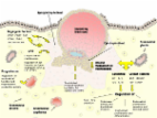
          Leptin (Lep) is known for its involvement in the regulation of reproductive functions. It is important for uterine receptivity, implantation, placental growth and maternal energy homeostasis in several species, but Lep’s function in the pregnant dog has not been investigated.

          Methods

          Pregnant bitches were ovariohysterectomized at pre-implantation, post-implantation, mid-gestation and prepartum luteolysis. Two additional groups were treated with aglepristone in mid-gestation, and ovariohysterectomized 24 or 72 h later. Lep and leptin receptor (LepR) gene expression was detected by semi-quantitative real-time PCR in pre-implantation and inter-placental uterine sections (Ut) and in utero-placental compartments (Ut/Pl). Immunohistochemistry and in situ hybridization (ISH) were performed for Lep and LepR protein and mRNA localization. Parametric one-way ANOVA, paired t-test and Wilcoxon signed-rank test were used for statistical analysis.

          Results

          In the Ut/Pl, Lep expression was higher at post-implantation and prepartum luteolysis than at mid-gestation, while in the Ut, Lep mRNA levels did not change during pregnancy. LepR expression in the Ut/Pl was up-regulated at prepartum luteolysis compared to the earlier stages. In the Ut, highest LepR mRNA was found at pre- and post-implantation. LepR expression was down-regulated in the Ut/Pl compared to the Ut at post-implantation and at mid-gestation. Aglepristone treatment resulted in a decrease of Lep mRNA levels from 24 to 72 h in the Ut without concomitant changes in the Ut/Pl or in LepR levels. Lep and LepR immunoreactivities were strong in the luminal and glandular epithelium in the Ut with abundant LepR signals in the subepithelial stroma. In the Ut/Pl, fetal trophoblasts stained stronger for Lep and LepR than decidual cells, and signals for both proteins were also detected in the glandular chambers. The myometrium, blood vessel media, and sporadically also the endothelium stained for Lep and LepR. ISH showed similar signal distribution in the Ut and Ut/Pl.

          Conclusions

          Lep and LepR are differentially expressed in the canine uterus and placenta during pregnancy, and their presence in various cell types indicates paracrine/autocrine roles. The Lep signaling system may be one of the pathways involved in feto-maternal cross-talk, implantation and maintenance of pregnancy, and may have a regulatory role around parturition.

          Related collections

          Most cited references72

          • Record: found
          • Abstract: found
          • Article: not found

          Intracellular signalling pathways activated by leptin.

          Leptin is a versatile 16 kDa peptide hormone, with a tertiary structure resembling that of members of the long-chain helical cytokine family. It is mainly produced by adipocytes in proportion to fat size stores, and was originally thought to act only as a satiety factor. However, the ubiquitous distribution of OB-R leptin receptors in almost all tissues underlies the pleiotropism of leptin. OB-Rs belong to the class I cytokine receptor family, which is known to act through JAKs (Janus kinases) and STATs (signal transducers and activators of transcription). The OB-R gene is alternatively spliced to produce at least five isoforms. The full-length isoform, OB-Rb, contains intracellular motifs required for activation of the JAK/STAT signal transduction pathway, and is considered to be the functional receptor. Considerable evidence for systemic effects of leptin on body mass control, reproduction, angiogenesis, immunity, wound healing, bone remodelling and cardiovascular function, as well as on specific metabolic pathways, indicates that leptin operates both directly and indirectly to orchestrate complex pathophysiological processes. Consistent with leptin's pleiotropic role, its participation in and crosstalk with some of the main signalling pathways, including those involving insulin receptor substrates, phosphoinositide 3-kinase, protein kinase B, protein kinase C, extracellular-signal-regulated kinase, mitogen-activated protein kinases, phosphodiesterase, phospholipase C and nitric oxide, has been observed. The impact of leptin on several equally relevant signalling pathways extends also to Rho family GTPases in relation to the actin cytoskeleton, production of reactive oxygen species, stimulation of prostaglandins, binding to diacylglycerol kinase and catecholamine secretion, among others.
            Bookmark
            • Record: found
            • Abstract: found
            • Article: not found

            Leptin induces vascular permeability and synergistically stimulates angiogenesis with FGF-2 and VEGF.

            Most endocrine hormones are produced in tissues and organs with permeable microvessels that may provide an excess of hormones to be transported by the blood circulation to the distal target organ. Here, we investigate whether leptin, an endocrine hormone, induces the formation of vascular fenestrations and permeability, and we characterize its angiogenic property in the presence of other angiogenic factors. We provide evidence that leptin-induced new blood vessels are fenestrated. Under physiological conditions, capillary fenestrations are found in the leptin-producing adipose tissue in lean mice. In contrast, no vascular fenestrations were detected in the adipose tissue of leptin-deficient ob/ob mice. Thus, leptin plays a critical role in the maintenance and regulation of vascular fenestrations in the adipose tissue. Leptin induces a rapid vascular permeability response when administrated intradermally. Further, leptin synergistically stimulates angiogenesis with fibroblast growth factor (FGF)-2 and vascular endothelial growth factor (VEGF), the two most potent and commonly expressed angiogenic factors. These findings demonstrate that leptin has another new function-the increase of vascular permeability.
              Bookmark
              • Record: found
              • Abstract: found
              • Article: found
              Is Open Access

              Human trophoblast function during the implantation process

              The implantation process involves complex and synchronized molecular and cellular events between the uterus and the implanting embryo. These events are regulated by paracrine and autocrine factors. Trophoblast invasion and migration through the uterine wall is mediated by molecular and cellular interactions, controlled by the trophoblast and the maternal microenvironment. This review is focused on the molecular constituents of the human trophoblast, their actions and interactions, including interrelations with the uterine endometrium.
                Bookmark

                Author and article information

                Contributors
                obalogh@vetclinics.uzh.ch
                lstaub@vetclinics.uzh.ch
                aykutgram@gmail.com
                boosa@vetanat.uzh.ch
                kowalewski@vetanat.uzh.ch
                ireichler@vetclinics.uzh.ch
                Journal
                Reprod Biol Endocrinol
                Reprod. Biol. Endocrinol
                Reproductive Biology and Endocrinology : RB&E
                BioMed Central (London )
                1477-7827
                8 March 2015
                8 March 2015
                2015
                : 13
                : 13
                Affiliations
                [ ]Clinic of Reproductive Medicine, Vetsuisse-Faculty, University of Zurich, Winterthurerstrasse 260, 8057 Zurich, Switzerland
                [ ]Institute of Veterinary Anatomy, Vetsuisse-Faculty, University of Zurich, Winterthurerstrasse 260, 8057 Zurich, Switzerland
                Article
                3
                10.1186/s12958-015-0003-6
                4358730
                25871422
                06ea72e9-a6f7-4325-8a9a-fb1c34fe0b73
                © Balogh et al.; licensee BioMed Central. 2015

                This is an Open Access article distributed under the terms of the Creative Commons Attribution License ( http://creativecommons.org/licenses/by/4.0), which permits unrestricted use, distribution, and reproduction in any medium, provided the original work is properly credited. The Creative Commons Public Domain Dedication waiver ( http://creativecommons.org/publicdomain/zero/1.0/) applies to the data made available in this article, unless otherwise stated.

                History
                : 6 October 2014
                : 6 February 2015
                Categories
                Research
                Custom metadata
                © The Author(s) 2015

                Human biology
                leptin,leptin receptor,dog,gestation,uterus,placenta,immunohistochemistry,in situ hybridization
                Human biology
                leptin, leptin receptor, dog, gestation, uterus, placenta, immunohistochemistry, in situ hybridization

                Comments

                Comment on this article