10
views
0
recommends
+1 Recommend
0 collections
    4
    shares
      • Record: found
      • Abstract: found
      • Article: not found

      Electrocardiogram in apical hypertrophic cardiomyopathy with a speculation as to the mechanism of its features

      research-article

      Read this article at

      ScienceOpenPublisherPMC
      Bookmark
          There is no author summary for this article yet. Authors can add summaries to their articles on ScienceOpen to make them more accessible to a non-specialist audience.

          Abstract

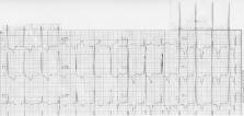
          A variety of electrocardiogram (ECG) alterations in patients with apical hypertrophic cardiomyopathy (AHCM) have been described in the literature, but no relevant quantitative analysis has been provided; thus the objective of this communication was to review the relevant literature and using two cases of patients with AHCM, to provide such a quantitative analysis. Using PubMed to search the literature 13 studies on the ECG in patients with AHCM were identified and evaluated; also a quantitative analysis of the ECG attributes in two patients was carried out. Qualitative ECG features from the literature on patients with AHCM is discussed. Also a description of the ECG in two patients with AHCM has identified as typical features truly giant QRS complexes in the precordial leads, particularly in lead V4, with rightward superior, and posterior shift of the T-wave vector, the latter being a newly described ECG correlate of AHCM. A speculation as to the possible mechanism of the observed ECG features is included.

          Related collections

          Most cited references14

          • Record: found
          • Abstract: found
          • Article: not found

          Disappearance of giant negative T waves in patients with the Japanese form of apical hypertrophy.

          The present study investigated the long-term changes in the electrocardiographic (ECG) hallmarks of the Japanese form of apical hypertrophy.
            Bookmark
            • Record: found
            • Abstract: found
            • Article: not found

            Apical hypertrophic cardiomyopathy might lead to misdiagnosis of ischaemic heart disease.

            In this study, demographic, clinic, electrocardiographic and angiographic properties of patients, on whom coronary angiography was performed with the pre-diagnosis of coronary artery disease (CAD) and whose ventriculography demonstrated typical apical hypertrophic cardiomyopathy (AHCM), were investigated. Seventeen patients (mean age 58 +/- 10 years, 10 male) with CAD pre-diagnosis, on whom coronary angiography was performed and had typical spade-like appearance on left ventriculography, were included in the study between January 2000 and May 2005. As risk factor for CAD, 8 (47%) patients had hypertension, 8 (47%) patients had dyslipidaemia, 2 (11%) patients had type 2 diabetes mellitus, 13 (77%) patients had a history of smoking, and 2 (11%) patients had family history. Seven (42%) patients presented unstable angina pectoris, 8 (47%) patients presented stable angina pectoris and 2 (11%) patients were asymptomatic. On coronary angiography, it was determined that 10 (58%) patients had normal coronary arteries, 3 (17%) patients had non-significant stenosis and 4 (25%) patients had myocardial bridging. Five (30%) patients revealed mid-ventricular obstruction and intraventricular gradient was 25 +/- 5 mmHg by the catheterization. All patients showed ''giant'' negative (> or = 10 mm) T waves in the precordial leads, whereas 2 patients had atrial fibrillation. Maximum wall thickness was measured as 18 +/- 4 mm in the apical region by transthoracic echocardiography. One patient (5%) who had mid-ventricular obstruction developed atrial fibrillation during 2 years follow-up, though any other events did not occur during hospitalization or follow-up period. Physicians caring for patients with chest pain should consider AHCM in their differential diagnosis in case of a patient with chest pain and electrocardiographic changes suggestive of CAD.
              Bookmark
              • Record: found
              • Abstract: found
              • Article: not found

              Long-term evaluation of patients with apical hypertrophic cardiomyopathy. Correlation between quantitative echocardiographic assessment of apical hypertrophy and clinical-electrocardiographic findings.

              Apical hypertrophic cardiomyopathy (AHCM) is characterized by primary hypertrophy localized exclusively in the apex of the left ventricle. Previous studies have indicated that AHCM results in a unique combination of cross-sectional echocardiographic (CSE) and ECG findings ('giant' T wave inversion and high R wave voltage in the precordial leads). The aims of this study were: (1) to assess the degree of AHCM in a quantitative fashion (2) to evaluate the possible relationship between apical hypertrophy, quantitatively determined, and ECG findings in patients with AHCM (3) to verify the changes in echocardiographic and ECG parameters over time (4) to define the relationship between the severity of AHCM and the clinical course of such patients. Eleven selected patients with AHCM were studied for an average 6 year follow-up period; there were seven men and four women (age from 18 to 62 years, mean 49). Apical hypertrophy was assessed quantitatively by determining the muscle cross-sectional area in the apical region, which was considered an index of myocardial mass. From the end-diastolic apical four chamber view, endocardial and epicardial contours were digitized in order to obtain the total muscle cross-sectional area of the left ventricle. The walls of the left ventricle were then divided into three regions (basal, intermediate, apical). The final value of each cross-sectional muscle area was obtained from the mean measurements of four independent and blinded observers. In AHCM the apical muscle cross-sectional area (AMA) ranged from 10.3 to 17.9 cm2, mean 13.2 +/- 2.6 cm2.(ABSTRACT TRUNCATED AT 250 WORDS)
                Bookmark

                Author and article information

                Contributors
                +1-718-3345005 , +1-718-3345990 , madiasj@nychhc.org
                Journal
                Neth Heart J
                Neth Heart J
                Netherlands Heart Journal
                Bohn Stafleu van Loghum (Heidelberg )
                1568-5888
                1876-6250
                18 May 2013
                18 May 2013
                June 2013
                : 21
                : 6
                : 268-271
                Affiliations
                [ ]Mount Sinai School of Medicine, New York University, New York, NY USA
                [ ]Division of Cardiology, Elmhurst Hospital Center, 79-01 Broadway, Elmhurst, NY 11373 USA
                Article
                400
                10.1007/s12471-013-0400-4
                3661871
                23686564
                07acbdb7-d0ea-42bc-8d7a-85089b710885
                © The Author(s) 2013

                Open Access This article is distributed under the terms of the Creative Commons Attribution License which permits any use, distribution, and reproduction in any medium, provided the original author(s) and the source are credited.

                History
                Categories
                Review Article
                Custom metadata
                © The Author(s) 2013

                Cardiovascular Medicine
                electrocardiogram,electrophysiology,hypertrophic cardiomyopathy,apical hypertrophic cardiomyopathy,giant r-waves,giant t-waves

                Comments

                Comment on this article