22
views
0
recommends
+1 Recommend
0 collections
    0
    shares
      • Record: found
      • Abstract: found
      • Article: found
      Is Open Access

      Prostate motion during radiotherapy of prostate cancer patients with and without application of a hydrogel spacer: a comparative study

      research-article

      Read this article at

      Bookmark
          There is no author summary for this article yet. Authors can add summaries to their articles on ScienceOpen to make them more accessible to a non-specialist audience.

          Abstract

          Background and purpose

          The use of a tissue expander (hydrogel) for sparing of the rectum from increased irradiation during prostate radiotherapy is becoming popular. The goal of this study is to investigate the effect of a tissue expander (hydrogel) on the intrafraction prostate motion during radiotherapy.

          Methods and material

          Real time prostate motion was analysed for 26 patients and 742 fractions; 12 patients with and 14 patients without hydrogel (SpaceOAR™). The intra-fraction motion was quantified and compared between the two groups.

          Results

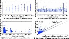
          The average (±standard deviation) of the mean motion during the treatment for patients with and without hydrogel was 1.5 (±0.8 mm) and 1.1 (±0.9 mm) respectively (p < 0.05). The average time of motion >3 mm for patients with and without hydrogel was 7.7 % (±1.1 %) and 4.5 % (±0.9 %) respectively (p > 0.05). The hydrogel age, fraction number and treatment time were found to have no effect ( R 2  < 0.05) on the prostate motion.

          Conclusions

          Differences in intrafraction motion in patients with hydrogel and without hydrogel were within measurement uncertainty (<1 mm). This result confirms that the addition of a spacer does not negate the need for intrafraction motion management if clinically indicated.

          Related collections

          Most cited references23

          • Record: found
          • Abstract: found
          • Article: not found
          Is Open Access

          Stereotactic body radiotherapy for localized prostate cancer: pooled analysis from a multi-institutional consortium of prospective phase II trials.

          The effectiveness of stereotactic body radiotherapy (SBRT) for localized prostate cancer is tested. A total of 1100 patients with clinically localized prostate cancer were enrolled in separate prospective phase 2 clinical trials of SBRT from 8 institutions during 2003-11 and pooled for analysis. SBRT using the CyberKnife delivered a median dose of 36.25Gy in 4-5 fractions. Patients were low-risk (58%), intermediate-risk (30%) and high-risk (11%). A short-course of androgen deprivation therapy (ADT) was given to 14%. PSA relapse defined as a rise >2ng/ml above nadir was analyzed with the Kaplan Meier method. With a median follow-up of 36months there were 49 patients with PSA failure (4.5%), 9 of whom were subsequently determined to be benign PSA bounces. The 5-year biochemical relapse free survival (bRFS) rate was 93% for all patients; 95%, 83% and 78% for GS ⩽6, 7 and ⩾8, respectively (p=0.001), and 95%, 84% and 81% for low-, intermediate- and high-risk patients, respectively (p 0.2ng/ml was noted among 16% of patients. For 135 patients possessing a minimum of 5years follow-up, the 5-year bRFS rate for low- and intermediate-risk patients was 99% and 93%, respectively. PSA relapse-free survival rates after SBRT compare favorably with other definitive treatments for low and intermediate risk patients. The current evidence supports consideration of SBRT among the therapeutic options for these patients. Copyright © 2013 The Authors. Published by Elsevier Ireland Ltd.. All rights reserved.
            Bookmark
            • Record: found
            • Abstract: found
            • Article: not found

            Preliminary toxicity analysis of 3-dimensional conformal radiation therapy versus intensity modulated radiation therapy on the high-dose arm of the Radiation Therapy Oncology Group 0126 prostate cancer trial.

            To give a preliminary report of clinical and treatment factors associated with toxicity in men receiving high-dose radiation therapy (RT) on a phase 3 dose-escalation trial. The trial was initiated with 3-dimensional conformal RT (3D-CRT) and amended after 1 year to allow intensity modulated RT (IMRT). Patients treated with 3D-CRT received 55.8 Gy to a planning target volume that included the prostate and seminal vesicles, then 23.4 Gy to prostate only. The IMRT patients were treated to the prostate and proximal seminal vesicles to 79.2 Gy. Common Toxicity Criteria, version 2.0, and Radiation Therapy Oncology Group/European Organization for Research and Treatment of Cancer late morbidity scores were used for acute and late effects. Of 763 patients randomized to the 79.2-Gy arm of Radiation Therapy Oncology Group 0126 protocol, 748 were eligible and evaluable: 491 and 257 were treated with 3D-CRT and IMRT, respectively. For both bladder and rectum, the volumes receiving 65, 70, and 75 Gy were significantly lower with IMRT (all P 15%) volumes of rectum >70 Gy are associated with late rectal toxicity. Copyright © 2013 Elsevier Inc. All rights reserved.
              Bookmark
              • Record: found
              • Abstract: found
              • Article: not found

              Observations on real-time prostate gland motion using electromagnetic tracking.

              To quantify and describe the real-time movement of the prostate gland in a large data set of patients treated with radiotherapy. The Calypso four-dimensional localization system was used for target localization in 17 patients, with electromagnetic markers implanted in the prostate of each patient. We analyzed a total of 550 continuous tracking sessions. The fraction of time that the prostate was displaced by >3, >5, >7, and >10 mm was calculated for each session and patient. The frequencies of displacements after initial patient positioning were analyzed over time. Averaged over all patients, the prostate was displaced >3 and >5 mm for 13.6% and 3.3% of the total treatment time, respectively. For individual patients, the corresponding maximal values were 36.2% and 10.9%. For individual fractions, the corresponding maximal values were 98.7% and 98.6%. Displacements >3 mm were observed at 5 min after initial alignment in about one-eighth of the observations, and increased to one-quarter by 10 min. For individual patients, the maximal value of the displacements >3 mm at 5 and 10 min after initial positioning was 43% and 75%, respectively. On average, the prostate was displaced by >3 mm and >5 mm approximately 14% and 3% of the time, respectively. For individual patients, these values were up to three times greater. After the initial positioning, the likelihood of displacement of the prostate gland increased with elapsed time. This highlights the importance of initiating treatment shortly after initially positioning the patient.
                Bookmark

                Author and article information

                Contributors
                +61 2 9351 8731 , prabhjot@physics.usyd.edu.au
                Andrew.Kneebone@health.nsw.gov.au
                Jeremy.Booth@health.nsw.gov.au
                david.thwaites@sydney.edu.au
                kaurramandeep@hotmail.co.uk
                emma.colvill@sydney.edu.au
                jin.aun@sydney.edu.au
                paul.keall@sydney.edu.au
                Thomas.Eade@health.nsw.gov.au
                Journal
                Radiat Oncol
                Radiat Oncol
                Radiation Oncology (London, England)
                BioMed Central (London )
                1748-717X
                24 October 2015
                24 October 2015
                2015
                : 10
                : 215
                Affiliations
                [ ]Northern Sydney Cancer Centre, Royal North Shore Hospital, Sydney, NSW 2065 Australia
                [ ]Institute of Medical Physics, School of Physics, University of Sydney, Sydney, NSW 2006 Australia
                [ ]5/161A Willoughby Road, Naremburn, NSW 2065 Australia
                [ ]Radiation Physics Laboratory, School of Medicine, University of Sydney, Sydney, NSW 2006 Australia
                Article
                526
                10.1186/s13014-015-0526-1
                4619294
                26499473
                0b44dc6a-f2bc-4834-89e6-fcd295fb3c53
                © Juneja et al. 2015

                Open AccessThis article is distributed under the terms of the Creative Commons Attribution 4.0 International License ( http://creativecommons.org/licenses/by/4.0/), which permits unrestricted use, distribution, and reproduction in any medium, provided you give appropriate credit to the original author(s) and the source, provide a link to the Creative Commons license, and indicate if changes were made. The Creative Commons Public Domain Dedication waiver ( http://creativecommons.org/publicdomain/zero/1.0/) applies to the data made available in this article, unless otherwise stated.

                History
                : 10 July 2015
                : 19 October 2015
                Categories
                Research
                Custom metadata
                © The Author(s) 2015

                Oncology & Radiotherapy
                prostate cancer,radiotherapy,intrafraction motion,hydrogel spacer
                Oncology & Radiotherapy
                prostate cancer, radiotherapy, intrafraction motion, hydrogel spacer

                Comments

                Comment on this article