104
views
0
recommends
+1 Recommend
0 collections
    0
    shares
      • Record: found
      • Abstract: found
      • Article: found
      Is Open Access

      Clinical Evidence for the Relationship between Nail Configuration and Mechanical Forces

      research-article
      , MD *† , , , MD, PhD
      Plastic and Reconstructive Surgery Global Open
      Wolters Kluwer Health

      Read this article at

      Bookmark
          There is no author summary for this article yet. Authors can add summaries to their articles on ScienceOpen to make them more accessible to a non-specialist audience.

          Abstract

          Summary:

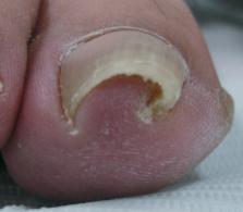
          Mechanobiology is an emerging field of science that focuses on the way physical forces and changes in cell or tissue mechanics contribute to development, physiology, and disease. As nails are always exposed to physical stimulation, mechanical forces may have a particularly pronounced effect on nail configuration and could be involved in the development of nail deformities. However, the role of mechanobiology in nail configuration and deformities has rarely been assessed. This review describes what is currently understood regarding the effect of mechanical force on nail configuration and deformities. On the basis of these observations, we hypothesize that nails have an automatic curvature function that allows them to adapt to the daily upward mechanical forces. Under normal conditions, the upward daily mechanical force and the automatic curvature force are well balanced. However, an imbalance between these 2 forces may cause nail deformation. For example, pincer nails may be caused by the absence of upward mechanical forces or a genetic propensity increase in the automatic curvature force, whereas koilonychias may occur when the upward mechanical force exceeds the automatic curvature force, thereby causing the nail to curve outward. This hypothesis is a new concept that could aid the development of innovative methods to prevent and treat nail deformities.

          Related collections

          Most cited references64

          • Record: found
          • Abstract: found
          • Article: not found

          Mechanotransduction and the functional response of bone to mechanical strain.

          Mechanotransduction plays a crucial role in the physiology of many tissues including bone. Mechanical loading can inhibit bone resorption and increase bone formation in vivo. In bone, the process of mechanotransduction can be divided into four distinct steps: (1) mechanocoupling, (2) biochemical coupling, (3) transmission of signal, and (4) effector cell response. In mechanocoupling, mechanical loads in vivo cause deformations in bone that stretch bone cells within and lining the bone matrix and create fluid movement within the canaliculae of bone. Dynamic loading, which is associated with extracellular fluid flow and the creation of streaming potentials within bone, is most effective for stimulating new bone formation in vivo. Bone cells in vitro are stimulated to produce second messengers when exposed to fluid flow or mechanical stretch. In biochemical coupling, the possible mechanisms for the coupling of cell-level mechanical signals into intracellular biochemical signals include force transduction through the integrin-cytoskeleton-nuclear matrix structure, stretch-activated cation channels within the cell membrane, G protein-dependent pathways, and linkage between the cytoskeleton and the phospholipase C or phospholipase A pathways. The tight interaction of each of these pathways would suggest that the entire cell is a mechanosensor and there are many different pathways available for the transduction of a mechanical signal. In the transmission of signal, osteoblasts, osteocytes, and bone lining cells may act as sensors of mechanical signals and may communicate the signal through cell processes connected by gap junctions. These cells also produce paracrine factors that may signal osteoprogenitors to differentiate into osteoblasts and attach to the bone surface. Insulin-like growth factors and prostaglandins are possible candidates for intermediaries in signal transduction. In the effector cell response, the effects of mechanical loading are dependent upon the magnitude, duration, and rate of the applied load. Longer duration, lower amplitude loading has the same effect on bone formation as loads with short duration and high amplitude. Loading must be cyclic to stimulate new bone formation. Aging greatly reduces the osteogenic effects of mechanical loading in vivo. Also, some hormones may interact with local mechanical signals to change the sensitivity of the sensor or effector cells to mechanical load.
            Bookmark
            • Record: found
            • Abstract: found
            • Article: not found

            Biology of the hair follicle: the basics.

            The mammalian hair follicle represents a unique, highly regenerative neuroectodermal-mesodermal interaction system that contains numerous stem cells. It is the only organ in the mammalian organism that undergoes life-long cycles of rapid growth (anagen), regression (catagen), and resting periods (telogen). These transformations are controlled by changes in the local signaling milieu, based on changes in expression/activity of a constantly growing number of cytokines, hormones, neurotransmitters, and their cognate receptors as well as of transcription factors and enzymes that have become recognized as key mediators of hair follicle cycling. Transplantation experiments have shown that the driving force of cycling, the "hair cycle clock," is located in the hair follicle itself. However, the exact underlying molecular mechanisms that drive this oscillator system remain unclear. These controls of hair follicle cycling are of great clinical interest because hair loss or unwanted hair growth largely reflect undesired changes in hair follicle cycling. To develop therapeutic agents for the management of these hair cycle abnormalities, it is critical to decipher and pharmacologically target the key molecular controls that underlie the enigmatic "hair cycle clock."
              Bookmark
              • Record: found
              • Abstract: found
              • Article: not found

              Mechanotherapy: revisiting physical therapy and recruiting mechanobiology for a new era in medicine.

              It has long been thought that the effectiveness and efficiency of physical therapy would improve if our understanding of the cell biology/biochemistry that participates in mechanics could be improved. Traditional physical therapy focuses primarily on rehabilitation, but recent developments in mechanobiology that illuminated the effects of physical forces on cells and tissues have led to the realization that the old therapy model should be updated. To achieve this here, the term mechanotherapy is proposed and recent studies showing how mechanotherapies target particular cells, molecules, and tissues are reviewed. These studies show how mechanical force modulates integrin-mediated processes and other mechanosensors such as gap junctions, hemichannels, primary cilia, transient receptor potential channels (cell targeting), and intracellular mechanosignaling pathways (molecule targeting). The role of mechanical force in various therapies, including microdeformation, shockwave, tissue expansion, distraction osteogenesis, and surgical tension reduction (tissue targeting) therapies, is reviewed. This review aims to jumpstart research into this field, which promises to generate a new era of viable and novel pharmacological and engineering interventions that can overcome human diseases. Copyright © 2013 Elsevier Ltd. All rights reserved.
                Bookmark

                Author and article information

                Journal
                Plast Reconstr Surg Glob Open
                Plast Reconstr Surg Glob Open
                GOX
                Plastic and Reconstructive Surgery Global Open
                Wolters Kluwer Health
                2169-7574
                March 2014
                07 April 2014
                : 2
                : 3
                : e115
                Affiliations
                From the [* ]Department of Surgical Science, Graduate School of Medicine, University of Tokyo, Tokyo, Japan; and []Department of Plastic, Reconstructive and Aesthetic Surgery, Nippon Medical School, Tokyo, Japan.
                Author notes
                Hitomi Sano, Department of Surgical Science Graduate School of Medicine University of Tokyo Hongo 7-3-1, Bunkyo-ku Tokyo 113–8656 Japan E-mail: sasasa116sasasa@ 123456hotmail.com
                Article
                00004
                10.1097/GOX.0000000000000057
                4174141
                25289309
                0b541e37-1ca7-412d-8a87-781da5dea6ef
                Copyright © 2013 The Authors. Published by Lippincott Williams & Wilkins on behalf of The American Society of Plastic Surgeons. PRS Global Open is a publication of the American Society of Plastic Surgeons.

                This is an open-access article distributed under the terms of the Creative Commons Attribution-NonCommercial-NoDerivitives 3.0 License, where it is permissible to download and share the work provided it is properly cited. The work cannot be changed in any way or used commercially.

                History
                : 14 August 2013
                : 02 January 2014
                Categories
                Original Articles
                Custom metadata
                TRUE

                Comments

                Comment on this article