68
views
0
recommends
+1 Recommend
0 collections
    0
    shares
      • Record: found
      • Abstract: found
      • Article: found
      Is Open Access

      Mutations that are a common cause of Leber congenital amaurosis in northern America are rare in Southern India

      research-article

      Read this article at

      Bookmark
          There is no author summary for this article yet. Authors can add summaries to their articles on ScienceOpen to make them more accessible to a non-specialist audience.

          Abstract

          Purpose

          To test patients from southern India for the presence of mutations that most commonly cause Leber congenital amaurosis (LCA) in northern America.

          Methods

          A review of the literature identified 177 unique LCA causing mutations in eight different genes: aryl hydrocarbon receptor interacting protein-like 1 ( AIPL1), crumbs homolog 1 ( CRB1), cone-rod homeobox ( CRX), guanylate cyclase 2D ( GUCY2D), nephronophthisis 6 ( NPHP6), retinol dehydrogenase 12 ( RDH12), retinal pigment epithelium-specific protein 65 kDa ( RPE65), and retinitis pigmentosa GTPase regulator interacting protein 1 ( RPGRIP1). Allele-specific ligation assay and bidirectional sequencing were used to test 38 unrelated LCA patients from southern India for 104 of these mutations, which contribute to more than 30% of the LCA cases in a northern American population.

          Results

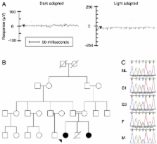
          Only one participant was found to harbor one of the 104 mutations in the allele-specific assay (homozygous RPE65 Tyr368His). A mutation that was not part of the assay (homozygous RPE65 Tyr143Asp) was incidentally detected in a second patient when an equivocal signal from one allele on the assay was followed up with automated DNA sequencing.

          Conclusions

          Mutations that contribute to 30% of the LCA cases in northern America were detected in only 2.6% of LCA cases in our cohort from southern India. There were no instances of IVS26 c.2991+1655 A>G in NPHP6, the most commonly detected mutation in LCA. These data suggest that LCA in India is caused primarily by a different set of mutations in the same genes associated with disease in northern America, or by mutations in other genes that have not yet been discovered. Therefore, mutation-specific assays developed for European and northern American cohorts may not be suited for testing LCA patients from India or other ethnically distinct populations.

          Related collections

          Most cited references15

          • Record: found
          • Abstract: found
          • Article: not found

          Mutations in RPE65 cause autosomal recessive childhood-onset severe retinal dystrophy.

          Autosomal recessive childhood-onset severe retinal dystrophy (arCSRD) designates a heterogeneous group of disorders affecting rod and cone photoreceptors simultaneously. The most severe cases are termed Leber congenital amaurosis (LCA), while the less aggressive forms are usually considered juvenile retinitis pigmentosa. Recently, mutations in the retinal-specific guanylate cyclase gene were found in patients with LCA. Disease genes implicated in other forms of arCSRD are expected to encode proteins present in the neuroretina or in the retinal pigment epithelium (RPE). The RPE, a monolayer of cells separating the vascular-rich choroid and the neuroretina, is in intimate contact with the outer segments of rods and cones via the microvilli surrounding the photoreceptors. The RPE expresses a tissue-specific and evolutionarily highly conserved 61 kD protein (RPE65) present at high levels in vivo. Although the function of RPE65 is not yet known, an important role in the RPE/photoreceptor vitamin-A cycle is suggested by the fact that RPE65 associates both with serum retinol-binding protein and with the RPE-specific 11-cis retinol dehydrogenase, an enzyme active in the synthesis of the visual pigment chromophore 11-cis retinal. Here we report that the analysis of RPE65 in a collection of about 100 unselected retinal-dystrophy patients of different ethnic origin revealed five that are likely to be pathogenic mutations, including a missense mutation (Pro363Thr), two point mutations affecting splicing (912 + 1G-->T and 65 + 5G-->A) and two small re-arrangements (ins144T and 831del8) on a total of nine alleles of five patients with arCSRD. In contrast to other genes whose defects have been implicated in degenerative retinopathies, RPE65 is the first disease gene in this group of inherited disorders that is expressed exclusively in the RPE, and may play a role in vitamin-A metabolism of the retina.
            Bookmark
            • Record: found
            • Abstract: found
            • Article: not found

            Mutations in a human homologue of Drosophila crumbs cause retinitis pigmentosa (RP12).

            Retinitis pigmentosa (RP) comprises a clinically and genetically heterogeneous group of diseases that afflicts approximately 1.5 million people worldwide. Affected individuals suffer from a progressive degeneration of the photoreceptors, eventually resulting in severe visual impairment. To isolate candidate genes for chorioretinal diseases, we cloned cDNAs specifically or preferentially expressed in the human retina and the retinal pigment epithelium (RPE) through a novel suppression subtractive hybridization (SSH) method. One of these cDNAs (RET3C11) mapped to chromosome 1q31-q32.1, a region harbouring a gene involved in a severe form of autosomal recessive RP characterized by a typical preservation of the para-arteriolar RPE (RP12; ref. 3). The full-length cDNA encodes an extracellular protein with 19 EGF-like domains, 3 laminin A G-like domains and a C-type lectin domain. This protein is homologous to the Drosophila melanogaster protein crumbs (CRB), and denoted CRB1 (crumbs homologue 1). In ten unrelated RP patients with preserved para-arteriolar RPE, we identified a homozygous AluY insertion disrupting the ORF, five homozygous missense mutations and four compound heterozygous mutations in CRB1. The similarity to CRB suggests a role for CRB1 in cell-cell interaction and possibly in the maintenance of cell polarity in the retina. The distinct RPE abnormalities observed in RP12 patients suggest that CRB1 mutations trigger a novel mechanism of photoreceptor degeneration.
              Bookmark
              • Record: found
              • Abstract: found
              • Article: not found

              Retinal-specific guanylate cyclase gene mutations in Leber's congenital amaurosis.

              Leber's congenital amaurosis (LCA, MIM 204,000), the earliest and most severe form of inherited retinopathy, accounts for at least 5% of all inherited retinal dystrophies. This autosomal recessive condition is usually recognized at birth or during the first months of life in an infant with total blindness or greatly impaired vision, normal fundus and extinguished electroretinogram (ERG). Nystagmus (pendular type) and characteristic eye poking are frequently observed in the first months of life (digito-ocular sign of Franceschetti). Hypermetropia and keratoconus frequently develop in the course of the disease. The observation by Waardenburg of normal children born to affected parents supports the genetic heterogeneity of LCA. Until now, however, little was known about the pathophysiology of the disease, but LCA is usually regarded as the consequence of either impaired development of photoreceptors or extremely early degeneration of cells that have developed normally. We have recently mapped a gene for LCA to chromosome 17p13.1 (LCA1) by homozygosity mapping in consanguineous families of North African origin and provided evidence of genetic heterogeneity in our sample, as LCA1 accounted for 8/15 LCA families in our series. Here, we report two missense mutations (F589S) and two frameshift mutations (nt 460 del C, nt 693 del C) of the retinal guanylate cyclase (RETGC, GDB symbol GUC2D) gene in four unrelated LCA1 probands of North African ancestry and ascribe LCA1 to an impaired production of cGMP in the retina, with permanent closure of cGMP-gated cation channels.
                Bookmark

                Author and article information

                Journal
                Mol Vis
                MV
                Molecular Vision
                Molecular Vision
                1090-0535
                2009
                04 September 2009
                : 15
                : 1781-1787
                Affiliations
                [1 ]Department of Genetics, Dr. G. Venkataswamy Eye Research Institute, Aravind Medical Research Foundation, Madurai, Tamilnadu, India
                [2 ]Department of Paediatric Ophthalmology, Aravind Eye Hospital, Madurai, Tamilnadu, India
                [3 ]Howard Hughes Medical Institute, Department of Ophthalmology and Visual Sciences, University of Iowa, Iowa City, IA
                [4 ]Department of Ophthalmology and Visual Sciences, University of Iowa, Iowa City, IA
                Author notes
                Correspondence to: Edwin M. Stone, Howard Hughes Medical Institute, Department of Ophthalmology and Visual Sciences, 4111 MERF, 375 Newton Road, University of Iowa, Iowa City, IA, 52246; Phone: (319) 594-8270; FAX: (319) 335-7142; email: edwin-stone@ 123456uiowa.edu
                Article
                188 2008MOLVIS0147
                2742639
                19753312
                0c497391-6bae-4f53-ad81-a4db8336a666
                Copyright @ 2009

                This is an open-access article distributed under the terms of the Creative Commons Attribution License, which permits unrestricted use, distribution, and reproduction in any medium, provided the original work is properly cited.

                History
                : 13 May 2008
                : 19 August 2009
                Categories
                Research Article
                Custom metadata
                Export to XML
                Stone

                Vision sciences
                Vision sciences

                Comments

                Comment on this article