19
views
0
recommends
+1 Recommend
0 collections
    0
    shares
      • Record: found
      • Abstract: found
      • Article: found
      Is Open Access

      Large gaze shift generation while standing: the role of the vestibular system

      research-article

      Read this article at

      Bookmark
          There is no author summary for this article yet. Authors can add summaries to their articles on ScienceOpen to make them more accessible to a non-specialist audience.

          Abstract

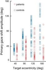
          The functional significance of vestibular information for the generation of gaze shifts is controversial and less well established than the vestibular contribution to gaze stability. In this study, we asked seven bilaterally avestibular patients to execute voluntary, whole body pivot turns to visual targets up to 180° while standing. In these conditions, not only are the demands imposed on gaze transfer mechanisms more challenging, but also neck proprioceptive input represents an inadequate source of head-in-space motion information. Patients’ body segment was slower and jerky. In the absence of visual feedback, gaze advanced in small steps, closely resembling normal multiple-step gaze-shift patterns, but as a consequence of the slow head motion, target acquisition was delayed. In ~25% of trials, however, patients moved faster but the velocity of prematurely emerging slow-phase compensatory eye movements remained lower than head-in-space velocity due to vestibuloocular failure. During these trials, therefore, gaze advanced toward the target without interruption but, again, taking longer than when normal controls use single-step gaze transfers. That is, even when patients attempted faster gaze shifts, exposing themselves to gaze instability, they acquired distant targets significantly later than controls. Thus, while patients are upright, loss of vestibular information disrupts not only gaze stability but also gaze transfers. The slow and ataxic head and trunk movements introduce significant foveation delays. These deficits explain patients’ symptoms during upright activities and show, for the first time, the clinical significance of losing the so-called “anticompensatory” (gaze shifting) function of the vestibuloocular reflex.

          NEW & NOTEWORTHY Previous studies in sitting avestibular patients concluded that gaze transfers are not substantially compromised. Still, clinicians know that patients are impeded (e.g., looking side to side before crossing a road). We show that during large gaze transfers while standing, vestibularly derived head velocity signals are critical for the mechanisms governing reorientation to distant targets and multisegmental coordination. Our findings go beyond the traditional role of the vestibular system in gaze stability, extending it to gaze transfers, as well.

          Related collections

          Most cited references28

          • Record: found
          • Abstract: found
          • Article: not found

          Footedness is a better predictor than is handedness of emotional lateralization.

          A tremendous amount of experimental work has attempted to identify reliable behavioural predictors of cerebral lateralization. Preferred handedness has been the most popular predictor, but some recent reports suggest that preferred footedness may serve as a more accurate predictor of functional laterality, especially in the left-handed population. The present study sought to test this claim by selectively recruiting individuals with either 'crossed' lateral preferences (right-handed and left-footed or left-handed and right-footed) or 'uncrossed' lateral preferences (right-handed and right-footed or left-handed and left-footed). Lateralization of emotional perception was assessed with two blocks of the dichotic Emotional Words Test (EWT), and lateral preference for both handedness and footedness was assessed using self-report questionnaires. Ear advantage on the dichotic task varied significantly with preferred foot (P=0.003), but not with preferred hand. Cerebral lateralization may be more related to footedness than to other lateral preferences.
            Bookmark
            • Record: found
            • Abstract: found
            • Article: not found

            Coordination of the eyes and head during visual orienting.

            Changing the direction of the line of sight is essential for the visual exploration of our environment. When the head does not move, re-orientation of the visual axis is accomplished with high velocity, conjugate movements of the eyes known as saccades. Our understanding of the neural mechanisms that control saccadic eye movements has advanced rapidly as specific hypotheses have been developed, evaluated and sometimes rejected on the basis of new observations. Constraints on new hypotheses and new tests of existing models have often arisen from the careful assessment of behavioral observations. The definition of the set of features (or rules) of saccadic eye movements was critical in the development of hypotheses of their neural control. When the head is free to move, changes in the direction of the line of sight can involve simultaneous saccadic eye movements and movements of the head. When the head moves in conjunction with the eyes to accomplish these shifts in gaze direction, the rules that helped define head-restrained saccadic eye movements are altered. For example, the slope relationship between duration and amplitude for saccadic eye movements is reversed (the slope is negative) during gaze shifts of similar amplitude initiated with the eyes in different orbital positions. Modifications to the hypotheses developed in head-restrained subjects may be needed to account for these new observations. This review briefly recounts features of head-restrained saccadic eye movements, and then describes some of the characteristics of coordinated eye-head movements that have led to development of new hypotheses describing the mechanisms of gaze shift control.
              Bookmark
              • Record: found
              • Abstract: found
              • Article: not found

              Gaze control in humans: eye-head coordination during orienting movements to targets within and beyond the oculomotor range.

              Gaze, the direction of the visual axis in space, is the sum of the eye position relative to the head (E) plus head position relative to space (H). In the old explanation, which we call the oculocentric motor strategy, of how a rapid orienting gaze shift is controlled, it is assumed that 1) a saccadic eye movement is programmed with an amplitude equal to the target's offset angle, 2) this eye movement is programmed without reference to whether a head movement is planned, 3) if the head turns simultaneously the saccade is reduced in size by an amount equal to the head's contribution, and 4) the saccade is attenuated by the vestibuloocular reflex (VOR) slow phase. Humans have an oculomotor range (OMR) of about +/- 55 degrees. The use of the oculocentric motor strategy to acquire targets lying beyond the OMR requires programming saccades that cannot be made physically. We have studied in normal human subjects rapid horizontal gaze shifts to visible and remembered targets situated within and beyond the OMR at offsets ranging from 30 to 160 degrees. Heads were attached to an apparatus that permitted short unexpected perturbations of the head trajectory. The acceleration and deceleration phases of the head perturbation could be timed to occur at different points in the eye movement. 4. Single-step rapid gaze shifts of all sizes up to at least 160 degrees (the limit studied) could be accomplished with the classic single-eye saccade and an accompanying saccadelike head movement. In gaze shifts less than approximately 45 degrees, when head motion was prevented totally by the brake, the eye attained the target. For larger target eccentricities the gaze shift was interrupted by the brake and the average eye saccade amplitude was approximately 45 degrees, well short of the OMR. Thus saccadic eye movement amplitude was neurally, not mechanically, limited. When the head's motion was not perturbed by the brake, the eye saccade amplitude was a function of head velocity: for a given target offset, the faster the head the smaller the saccade. For gaze shifts to targets beyond the OMR and when head velocity was low, the eye frequently attained the 45 degrees position limit and remained there, immobile, until gaze attained the target.(ABSTRACT TRUNCATED AT 400 WORDS)
                Bookmark

                Author and article information

                Journal
                J Neurophysiol
                J. Neurophysiol
                jn
                J Neurophysiol
                JN
                Journal of Neurophysiology
                American Physiological Society (Bethesda, MD )
                0022-3077
                1522-1598
                1 November 2019
                4 September 2019
                4 September 2019
                : 122
                : 5
                : 1928-1936
                Affiliations
                [1] 1Department of Neurology, University of Ioannina , Ioannina, Greece
                [2] 2Akutnahe Rehabilitation, Kantonsspital Baden, Baden, Switzerland
                [3] 3Department of Speech and Language Therapy, University of Ioannina , Ioannina, Greece
                [4] 4Department of Brain Sciences (Neuro-otology Unit), Imperial College London, Charing Cross Hospital , London, United Kingdom
                Author notes
                Address for reprint requests and other correspondence: D. Anastasopoulos, Akutnahe Rehabilitation, Kantonsspital Baden, 5404 Baden, Switzerland (e-mail: danastas@ 123456nurs.uoa.gr ).
                Author information
                https://orcid.org/0000-0003-2777-7031
                Article
                JN-00343-2019 JN-00343-2019
                10.1152/jn.00343.2019
                6879955
                31483710
                0d8ac0a2-046d-4f24-abfe-9fd2c9c566c8
                Copyright © 2019 the American Physiological Society

                Licensed under Creative Commons Attribution CC-BY 4.0: © the American Physiological Society.

                History
                : 3 June 2019
                : 15 August 2019
                : 4 September 2019
                Funding
                Funded by: Medical Research Council (MRC) 10.13039/501100000265
                Award ID: MR/J004685/1
                Categories
                Research Article
                Control of Movement

                Neurology
                anticompensatory,bilateral vestibular loss,coordination,gaze,multisegmental,turns
                Neurology
                anticompensatory, bilateral vestibular loss, coordination, gaze, multisegmental, turns

                Comments

                Comment on this article