41
views
0
recommends
+1 Recommend
0 collections
    0
    shares
      • Record: found
      • Abstract: found
      • Article: found
      Is Open Access

      Piezo1 links mechanical forces to red blood cell volume

      research-article

      Read this article at

      Bookmark
          There is no author summary for this article yet. Authors can add summaries to their articles on ScienceOpen to make them more accessible to a non-specialist audience.

          Abstract

          Red blood cells (RBCs) experience significant mechanical forces while recirculating, but the consequences of these forces are not fully understood. Recent work has shown that gain-of-function mutations in mechanically activated Piezo1 cation channels are associated with the dehydrating RBC disease xerocytosis, implicating a role of mechanotransduction in RBC volume regulation. However, the mechanisms by which these mutations result in RBC dehydration are unknown. In this study, we show that RBCs exhibit robust calcium entry in response to mechanical stretch and that this entry is dependent on Piezo1 expression. Furthermore, RBCs from blood-cell-specific Piezo1 conditional knockout mice are overhydrated and exhibit increased fragility both in vitro and in vivo. Finally, we show that Yoda1, a chemical activator of Piezo1, causes calcium influx and subsequent dehydration of RBCs via downstream activation of the KCa3.1 Gardos channel, directly implicating Piezo1 signaling in RBC volume control. Therefore, mechanically activated Piezo1 plays an essential role in RBC volume homeostasis.

          DOI: http://dx.doi.org/10.7554/eLife.07370.001

          eLife digest

          Within our bodies, cells and tissues are constantly being pushed and pulled by their surrounding environment. These mechanical forces are then transformed into electrical or chemical signals by cells. This process is crucial for many biological structures, such as blood vessels, to develop correctly, and is also a key part of our senses of touch and hearing.

          In 2010, researchers discovered a group of ion channels—proteins embedded in the membrane that surrounds a cell—that open up when a force is applied and allow calcium and other ions to enter the cell. This movement of ions generates the electrical response of the cell to the applied force. However, not much is known about the roles of these ‘Piezo’ ion channels.

          Red blood cells experience significant forces when they pass through narrow blood vessels. In a disease called xerocytosis, the red blood cells become severely dehydrated and shrink. In 2013, researchers discovered that patients with this disease have mutations in the gene that codes for the Piezo1 protein: a Piezo protein that has also been linked to a role in blood vessel development in embryos. This suggested that Piezo1 may regulate the volume of red blood cells.

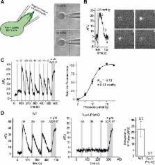
          Cahalan, Lukacs et al.—including some of the researchers who worked on the 2010 and 2013 studies—have now investigated the role of Piezo1 in red blood cells in more detail. Applying strong forces to red blood cells from mice caused calcium to rapidly enter cells through Piezo1 channels.

          Cahalan, Lukacs et al. then deleted the Piezo1 gene from red blood cells. This made the cells larger and more fragile than normal cells because they contained too much water. To investigate how Piezo1 regulates water content, the cells were treated with a chemical compound called Yoda1. This compound was shown in a separate study by Syeda et al. to activate Piezo1 channels. Activating Piezo1 caused a second type of ion channel to open up as well, which allowed potassium ions and water molecules to leave the cell. This resulted in the cell becoming dehydrated.

          This work raises the possibility that Piezo proteins are involved in other diseases where red blood cell volume is altered. In particular, many believe that Piezo1 may be involved in sickle cell disease, a possibility that can now be tested using the tools described in this study.

          DOI: http://dx.doi.org/10.7554/eLife.07370.002

          Related collections

          Most cited references26

          • Record: found
          • Abstract: found
          • Article: not found

          Piezos are pore-forming subunits of mechanically activated channels

          Mechanotransduction plays a crucial role in physiology. Biological processes including sensing touch and sound waves require yet unidentified cation channels that detect pressure. Mouse piezo1 (mpiezo1) and mpiezo2 induce mechanically activated cationic currents in cells; however, it is unknown if piezos are pore-forming ion channels or modulate ion channels. We show that Drosophila piezo (dpiezo) also induces mechanically activated currents in cells, but through channels with remarkably distinct pore properties including sensitivity to the pore blocker ruthenium red and single channel conductances. mpiezo1 assembles as a ~1.2 million-Dalton tetramer, with no evidence of other proteins in this complex. Finally, purified mpiezo1 reconstituted into asymmetric lipid bilayers and liposomes forms ruthenium red-sensitive ion channels. These data demonstrate that piezos are an evolutionarily conserved ion channel family involved in mechanotransduction.
            Bookmark
            • Record: found
            • Abstract: found
            • Article: not found

            Piezo1, a mechanically activated ion channel, is required for vascular development in mice.

            Mechanosensation is perhaps the last sensory modality not understood at the molecular level. Ion channels that sense mechanical force are postulated to play critical roles in a variety of biological processes including sensing touch/pain (somatosensation), sound (hearing), and shear stress (cardiovascular physiology); however, the identity of these ion channels has remained elusive. We previously identified Piezo1 and Piezo2 as mechanically activated cation channels that are expressed in many mechanosensitive cell types. Here, we show that Piezo1 is expressed in endothelial cells of developing blood vessels in mice. Piezo1-deficient embryos die at midgestation with defects in vascular remodeling, a process critically influenced by blood flow. We demonstrate that Piezo1 is activated by shear stress, the major type of mechanical force experienced by endothelial cells in response to blood flow. Furthermore, loss of Piezo1 in endothelial cells leads to deficits in stress fiber and cellular orientation in response to shear stress, linking Piezo1 mechanotransduction to regulation of cell morphology. These findings highlight an essential role of mammalian Piezo1 in vascular development during embryonic development.
              Bookmark
              • Record: found
              • Abstract: found
              • Article: found
              Is Open Access

              Chemical activation of the mechanotransduction channel Piezo1

              Piezo ion channels are activated by various types of mechanical stimuli and function as biological pressure sensors in both vertebrates and invertebrates. To date, mechanical stimuli are the only means to activate Piezo ion channels and whether other modes of activation exist is not known. In this study, we screened ∼3.25 million compounds using a cell-based fluorescence assay and identified a synthetic small molecule we termed Yoda1 that acts as an agonist for both human and mouse Piezo1. Functional studies in cells revealed that Yoda1 affects the sensitivity and the inactivation kinetics of mechanically induced responses. Characterization of Yoda1 in artificial droplet lipid bilayers showed that Yoda1 activates purified Piezo1 channels in the absence of other cellular components. Our studies demonstrate that Piezo1 is amenable to chemical activation and raise the possibility that endogenous Piezo1 agonists might exist. Yoda1 will serve as a key tool compound to study Piezo1 regulation and function. DOI: http://dx.doi.org/10.7554/eLife.07369.001
                Bookmark

                Author and article information

                Contributors
                Role: Reviewing editor
                Journal
                eLife
                eLife
                eLife
                eLife
                eLife Sciences Publications, Ltd
                2050-084X
                2050-084X
                22 May 2015
                2015
                : 4
                : e07370
                Affiliations
                [1 ]deptDepartment of Molecular and Cellular Neuroscience , Howard Hughes Medical Institute, The Scripps Research Institute , La Jolla, United States
                [2 ]deptDepartment of Bioengineering , University of California, San Diego , San Diego, United States
                [3 ]deptInstitute of Engineering in Medicine , University of California, San Diego , San Diego, United States
                [4 ]Genomics Institute of the Novartis Research Foundation , San Diego, United States
                Howard Hughes Medical Institute, Johns Hopkins University School of Medicine , United States
                Howard Hughes Medical Institute, Johns Hopkins University School of Medicine , United States
                Author notes
                [* ]For correspondence: ardem@ 123456scripps.edu
                [†]

                These authors contributed equally to this work.

                Article
                07370
                10.7554/eLife.07370
                4456639
                26001274
                0e2c41fd-681c-4574-b854-e197c170a6e0
                © 2015, Cahalan et al

                This article is distributed under the terms of the Creative Commons Attribution License, which permits unrestricted use and redistribution provided that the original author and source are credited.

                History
                : 07 March 2015
                : 08 May 2015
                Funding
                Funded by: FundRef http://dx.doi.org/10.13039/100000002, universityNational Institutes of Health (NIH);
                Award ID: NS083174
                Award Recipient :
                Funded by: FundRef http://dx.doi.org/10.13039/100000011, universityHoward Hughes Medical Institute (HHMI);
                Award ID: Investigator
                Award Recipient :
                Funded by: FundRef http://dx.doi.org/10.13039/100000900, universityCalifornia Institute for Regenerative Medicine (CIRM);
                Award Recipient :
                Funded by: FundRef http://dx.doi.org/10.13039/100000968, American Heart Association (AHA);
                Award ID: Postdoctoral Fellowship 14POST20000016
                Award Recipient :
                The funders had no role in study design, data collection and interpretation, or the decision to submit the work for publication.
                Categories
                Short Report
                Cell Biology
                Neuroscience
                Custom metadata
                2.3
                In red blood cells, the Piezo1 protein is responsible for sensing mechanical forces and volume regulation.

                Life sciences
                red blood cells,mechanotransduction,physiology,cell volume regulation,knockout animals,mouse

                Comments

                Comment on this article