19
views
0
recommends
+1 Recommend
0 collections
    0
    shares
      • Record: found
      • Abstract: found
      • Article: found
      Is Open Access

      Quantitative CT assessment of bone mineral density in dogs with hyperadrenocorticism

      research-article

      Read this article at

      Bookmark
          There is no author summary for this article yet. Authors can add summaries to their articles on ScienceOpen to make them more accessible to a non-specialist audience.

          Abstract

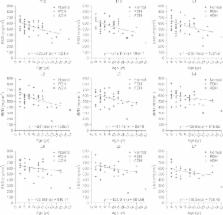
          Canine hyperadrenocorticism (HAC) is one of the most common causes of general osteopenia. In this study, quantitative computed tomography (QCT) was used to compare the bone mineral densities (BMD) between 39 normal dogs and 8 dogs with HAC (6 pituitary-dependent hyperadrenocorticism [PDH]; pituitary dependent hyperadrenocorticism, 2 adrenal hyperadrenocorticism [ADH]; adrenal dependent hyperadrenocorticism) diagnosed through hormonal assay. A computed tomogaraphy scan of the 12th thoracic to 7th lumbar vertebra was performed and the region of interest was drawn in each trabecular and cortical bone. Mean Hounsfield unit values were converted to equivalent BMD with bone-density phantom by linear regression analysis. The converted mean trabecular BMDs were significantly lower than those of normal dogs. ADH dogs showed significantly lower BMDs at cortical bone than normal dogs. Mean trabecular BMDs of dogs with PDH using QCT were significantly lower than those of normal dogs, and both mean trabecular and cortical BMDs in dogs with ADH were significantly lower than those of normal dogs. Taken together, these findings indicate that QCT is useful to assess BMD in dogs with HAC.

          Related collections

          Most cited references38

          • Record: found
          • Abstract: found
          • Article: not found

          Pre-existing fractures and bone mass predict vertebral fracture incidence in women.

          To determine the independent contributions of bone mass and existing fractures as predictors of the risk for new vertebral fractures. Postmenopausal Japanese-American women. Baseline measurements of the distal radius, the proximal radius, and the calcaneus were obtained in 1981 using single-photon absorptiometry. Measurements of the lumbar spine were obtained in 1984 using dual-photon absorptiometry. Prevalent vertebral fractures were identified using dimensions measured on lateral radiographs; vertebral height values more than 3 SD below vertebra-specific means were considered to indicate fracture. Statistical models were used to evaluate the utility of bone mass and existing (prevalent) fractures to predict the risk for new fractures during an average follow-up of 4.7 years. Differences of 2 SD in bone mass were associated with fourfold to sixfold increases in the risk for new vertebral fractures. A single fracture at the baseline examination increased the risk for new vertebral fractures fivefold. Presence of two or more fractures at baseline increased the risk 12-fold. A combination of low bone mass (below the 33d percentile) and the presence of two or more prevalent fractures increased the risk 75-fold, relative to women with the highest bone mass (above the 67th percentile) and no prevalent fractures. Stature, body mass index, arm span, and spinal conditions such as scoliosis, osteoarthritis, and sacroiliitis did not predict fracture incidence (P greater than 0.05). Weight was marginally predictive (P = 0.04) of fracture incidence but became nonpredictive after adjusting for bone mass (P greater than or equal to 0.05). Both bone mass and prevalent vertebral fractures are powerful predictors of the risk for new vertebral fractures. Combining information about bone mass and prevalent fracture appears to be better for predicting new fractures than either variable alone. Physicians can use these risk factors to identify patients at greatest risk for new fractures.
            Bookmark
            • Record: found
            • Abstract: found
            • Article: not found

            Precise measurement of vertebral mineral content using computed tomography.

            A precise method of measuring vertebral mineral content by computed tomography has been developed. Calibration phantoms scanned with patients are used to provide corrections for machine drifts. Advanced software manipulation of the display images is used to reposition precisely to the midportion of each vertebral body. A long-term precision of 2.8% for mineral content has been obtained in excised vertebrae in a phantom simulating the human torso.
              Bookmark
              • Record: found
              • Abstract: found
              • Article: not found

              Cushing's syndrome and bone.

              Structural and functional impairment of skeletal system is a relevant cause of morbidity and disability in patients with Cushing's syndrome (CS). Approximately 30-50% of patients with CS experience fractures (particularly at the spinal level) consistent with the 50% incidence of osteoporosis. Growth failure, pubertal arrest are the hallmarks of CS in children and growing adolescents leading to reduced final adult height and peak bone mass. The decrease in osteoblast number and function, through different mechanisms, seems to play a central role in the bone loss in CS. Patients with CS have decreased serum levels of osteocalcin and alkaline phosphatase. Considering the preferential bone loss in the cancellous skeleton it is reasonable to measure BMD, possibly with Dual X-rays absorptiometry (DEXA) at lumbar spine, in all patients with CS. Patients cured from CS have increased prevalence of spine damage: therefore, a radiological follow-up of the skeleton should be included in the management of patients with CS not only during the active phase but also after cure. Glucocorticoid-induced osteoporosis is reversible. The recovery of bone loss in CS is slow, taking approximately ten years to become complete. In the meanwhile, patients with severe osteopenia are exposed to a high risk of fracture. Alendronate may induce a more rapid improvement in BMD than cortisol normalization alone and it could be useful in patients with persistent postsurgical hypercortisolism to prevent further bone loss. The decision to discontinue antiresorptive therapy should be based on clinical monitoring and DEXA measurements.
                Bookmark

                Author and article information

                Journal
                J Vet Sci
                J. Vet. Sci
                JVS
                Journal of Veterinary Science
                The Korean Society of Veterinary Science
                1229-845X
                1976-555X
                December 2015
                17 December 2015
                : 16
                : 4
                : 531-542
                Affiliations
                [1 ]Department of Medical Imaging, College of Veterinary Medicine, Chungbuk National University, Cheongju 28644, Korea.
                [2 ]Laboratory of Internal Medicine, College of Veterinary Medicine, Chungbuk National University, Cheongju 28644, Korea.
                [3 ]Laboratory of Clinical Pathology, College of Veterinary Medicine, Chungbuk National University, Cheongju 28644, Korea.
                Author notes
                Corresponding author: Tel: +82-43-261-2609, Fax: +82-43-261-3224, dwchang@ 123456cbnu.ac.kr
                Article
                10.4142/jvs.2015.16.4.531
                4701747
                26040613
                114b9de5-05e4-4539-9cff-ffad1a510edb
                © 2015 The Korean Society of Veterinary Science.

                This is an Open Access article distributed under the terms of the Creative Commons Attribution Non-Commercial License ( http://creativecommons.org/licenses/by-nc/4.0) which permits unrestricted non-commercial use, distribution, and reproduction in any medium, provided the original work is properly cited.

                History
                : 19 November 2014
                : 10 March 2015
                : 04 April 2015
                Funding
                Funded by: National Research Foundation of Korea, CrossRef http://dx.doi.org/10.13039/501100003725;
                Award ID: NRF-2013R1A2A2A04008751
                Categories
                Original Article

                Veterinary medicine
                bone mineral density,dog,hyperadrenocorticism,quantitative computed tomography

                Comments

                Comment on this article