5
views
0
recommends
+1 Recommend
0 collections
    0
    shares
      • Record: found
      • Abstract: found
      • Article: found
      Is Open Access

      Cardiomyocytes Derived from Human CardiopoieticAmniotic Fluids

      research-article

      Read this article at

      Bookmark
          There is no author summary for this article yet. Authors can add summaries to their articles on ScienceOpen to make them more accessible to a non-specialist audience.

          Abstract

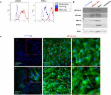
          Human amniotic fluid (hAF) cells share characteristics of both embryonic and adult stem cells. They proliferate rapidly and can differentiate into cells of all embryonic germ layers but do not form teratomas. Embryoid-bodies obtained from hAF have cardiac differentiation potential, but terminal differentiation to cardiomyocytes (CMs) has not yet been described. Our purpose was to promote cardiac differentiation in hAFcells. Cells were exposed to inducing factors for up to 15 days. Only the subset of hAF cells expressing the multipotency markers SSEA4, OCT4 and CD90 ( CardiopoieticAF cells) responded to the differentiation process by increasing the expression of the cardiac transcription factors Nkx2.5 and GATA4, sarcomeric proteins (cTnT, α-MHC, α-SA), Connexin43 and atrial and ventricular markers. Furthermore, differentiated cells were positive for the calcium pumps CACNA1C and SERCA2a, with approximately 30% of CardiopoieticAF-derived CM-like cells responding to caffeine or adrenergic stimulation. Some spontaneous rare beating foci were also observed. In conclusion, we demonstrated that CardiopoieticAF cells might differentiate toward the cardiac lineage giving rise to CM-like cells characterized by several cardiac-specific molecular, structural, and functional properties.

          Related collections

          Most cited references31

          • Record: found
          • Abstract: found
          • Article: not found

          hESC-Derived Cardiomyocytes Electrically Couple and Suppress Arrhythmias in Injured Hearts

          Transplantation studies in mice and rats have shown that human embryonic stem cell-derived cardiomyocytes (hESC-CMs) can improve the function of infarcted hearts 1–3 , but two critical issues related to their electrophysiological behavior in vivo remain unresolved. First, the risk of arrhythmias following hESC-CM transplantation in injured hearts has not been determined. Second, the electromechanical integration of hESC-CMs in injured hearts has not been demonstrated, so it is unclear if these cells improve contractile function directly through addition of new force-generating units. Here we use a guinea pig model to show hESC-CM grafts in injured hearts protect against arrhythmias and can contract synchronously with host muscle. Injured hearts with hESC-CM grafts show improved mechanical function and a significantly reduced incidence of both spontaneous and induced ventricular tachycardia (VT). To assess the activity of hESC-CM grafts in vivo, we transplanted hESC-CMs expressing the genetically-encoded calcium sensor, GCaMP3 4, 5 . By correlating the GCaMP3 fluorescent signal with the host ECG, we found that grafts in uninjured hearts have consistent 1:1 host-graft coupling. Grafts in injured hearts are more heterogeneous and typically include both coupled and uncoupled regions. Thus, human myocardial grafts meet physiological criteria for true heart regeneration, providing support for the continued development of hESC-based cardiac therapies for both mechanical and electrical repair.
            Bookmark
            • Record: found
            • Abstract: found
            • Article: not found

            Teratoma formation by human embryonic stem cells: evaluation of essential parameters for future safety studies.

            Transplantation of human embryonic stem cells (hESC) into immune-deficient mice leads to the formation of differentiated tumors comprising all three germ layers, resembling spontaneous human teratomas. Teratoma assays are considered the gold standard for demonstrating differentiation potential of pluripotent hESC and hold promise as a standard for assessing safety among hESC-derived cell populations intended for therapeutic applications. We tested the potency of teratoma formation in seven anatomical transplantation locations (kidney capsule, muscle, subcutaneous space, peritoneal cavity, testis, liver, epididymal fat pad) in SCID mice with and without addition of Matrigel, and found that intramuscular teratoma formation was the most experimentally convenient, reproducible, and quantifiable. In the same experimental setting, we compared undifferentiated hESC and differentiated populations enriched for either beating cardiomyocytes or definitive endoderm derivatives (insulin-secreting beta cells), and showed that all cell preparations rapidly formed teratomas with varying percentages of mesoderm, ectoderm, and endoderm. In limiting dilution experiments, we found that as little as two hESC colonies spiked into feeder fibroblasts produced a teratoma, while a more rigorous single-cell titration achieved a detection limit of 1/4000. In summary, we established core parameters essential for facilitating safety profiling of hESC-derived products for future therapeutic applications.
              Bookmark
              • Record: found
              • Abstract: found
              • Article: not found

              Induced Pluripotent Stem Cells 10 Years Later: For Cardiac Applications.

              Induced pluripotent stem cells (iPSCs) are reprogrammed cells that have features similar to embryonic stem cells, such as the capacity of self-renewal and differentiation into many types of cells, including cardiac myocytes. Although initially the reprogramming efficiency was low, several improvements in reprogramming methods have achieved robust and efficient generation of iPSCs without genomic insertion of transgenes. iPSCs display clonal variations in epigenetic and genomic profiles and cellular behavior in differentiation. iPSC-derived cardiac myocytes (iPSC cardiac myocytes) recapitulate phenotypic differences caused by genetic variations, making them attractive human disease models, and are useful for drug discovery and toxicology testing. In addition, iPSC cardiac myocytes can help with patient stratification in regard to drug responsiveness. Furthermore, they can be used as source cells for cardiac regeneration in animal models. Here, we review recent progress in iPSC technology and its applications to cardiac diseases.
                Bookmark

                Author and article information

                Contributors
                b.ghinassi@unich.it
                Journal
                Sci Rep
                Sci Rep
                Scientific Reports
                Nature Publishing Group UK (London )
                2045-2322
                13 August 2018
                13 August 2018
                2018
                : 8
                : 12028
                Affiliations
                [1 ]ISNI 0000 0001 2181 4941, GRID grid.412451.7, Department of Medicine and Aging Sciences, , University “G. d’Annunzio” of Chieti-Pescara, ; Via dei Vestini 31, 66100 Chieti, Italy
                [2 ]ISNI 0000 0001 2181 4941, GRID grid.412451.7, Department of Neuroscience, , Imaging e Clinical Scienes, University “G. d’Annunzio” of Chieti-Pescara, ; Via dei Vestini 31, 66100 Chieti, Italy
                [3 ]ISNI 0000 0001 2181 4941, GRID grid.412451.7, Department of Department of Psychological, Humanities and Territorial Sciences, , University “G. d’Annunzio” of Chieti-Pescara, ; Via dei Vestini 31, 66100 Chieti, Italy
                [4 ]ISNI 0000 0001 2285 2675, GRID grid.239585.0, Stem Cell Core Facility, , Columbia University Medical Center, ; 650 W. 168th St., 10032 New York, NY USA
                Author information
                http://orcid.org/0000-0002-4473-4909
                http://orcid.org/0000-0002-3529-2790
                Article
                30537
                10.1038/s41598-018-30537-z
                6089907
                30104705
                11684877-318c-4b92-8344-5f52705e058c
                © The Author(s) 2018

                Open Access This article is licensed under a Creative Commons Attribution 4.0 International License, which permits use, sharing, adaptation, distribution and reproduction in any medium or format, as long as you give appropriate credit to the original author(s) and the source, provide a link to the Creative Commons license, and indicate if changes were made. The images or other third party material in this article are included in the article’s Creative Commons license, unless indicated otherwise in a credit line to the material. If material is not included in the article’s Creative Commons license and your intended use is not permitted by statutory regulation or exceeds the permitted use, you will need to obtain permission directly from the copyright holder. To view a copy of this license, visit http://creativecommons.org/licenses/by/4.0/.

                History
                : 8 December 2017
                : 1 August 2018
                Funding
                Funded by: FundRef https://doi.org/10.13039/501100003407, Ministero dell'Istruzione, dell'Università e della Ricerca (Ministry of Education, University and Research);
                Award ID: SIR2014-RBSI140GLQ
                Award Recipient :
                Categories
                Article
                Custom metadata
                © The Author(s) 2018

                Uncategorized
                Uncategorized

                Comments

                Comment on this article