33
views
0
recommends
+1 Recommend
0 collections
    0
    shares
      • Record: found
      • Abstract: found
      • Article: not found

      Arrhythmogenic mechanisms in the isolated perfused hypokalaemic murine heart

      research-article

      Read this article at

      ScienceOpenPublisherPMC
      Bookmark
          There is no author summary for this article yet. Authors can add summaries to their articles on ScienceOpen to make them more accessible to a non-specialist audience.

          Abstract

          Aim

          Hypokalaemia is associated with a lethal form of ventricular tachycardia (VT), torsade de pointes, through pathophysiological mechanisms requiring clarification.

          Methods

          Left ventricular endocardial and epicardial monophasic action potentials were compared in isolated mouse hearts paced from the right ventricular epicardium perfused with hypokalaemic (3 and 4 m m [K +] o) solutions. Corresponding K + currents were compared in whole-cell patch-clamped epicardial and endocardial myocytes.

          Results

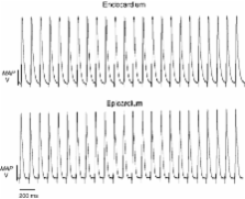
          Hypokalaemia prolonged epicardial action potential durations (APD) from mean APD 90s of 37.2 ± 1.7 ms ( n = 7) to 58.4 ± 4.1 ms ( n =7) and 66.7 ± 2.1 ms ( n = 11) at 5.2, 4 and 3 m m [K +] o respectively. Endocardial APD 90s correspondingly increased from 51.6 ± 1.9 ms ( n = 7) to 62.8 ± 2.8 ms ( n = 7) and 62.9 ± 5.9 ms ( n = 11) giving reductions in endocardial–epicardial differences, ΔAPD 90, from 14.4 ± 2.6 to 4.4 ± 5.0 and −3.4 ± 6.0 ms respectively. Early afterdepolarizations (EADs) occurred in epicardia in three of seven spontaneously beating hearts at 4 m m [K +] o with triggered beats followed by episodes of non-sustained VT in nine of 11 preparations at 3 m m. Programmed electrical stimulation never induced arrhythmic events in preparations perfused with normokalemic solutions yet induced VT in two of seven and nine of 11 preparations at 4 and 3 m m [K +] o respectively. Early outward K + current correspondingly fell from 73.46 ± 8.45 to 61.16±6.14 pA/pF in isolated epicardial but not endocardial myocytes ( n = 9) (3 m m [K +] o).

          Conclusions

          Hypokalaemic mouse hearts recapitulate the clinical arrhythmogenic phenotype, demonstrating EADs and triggered beats that might initiate VT on the one hand and reduced transmural dispersion of repolarization reflected in ΔAPD 90 suggesting arrhythmogenic substrate on the other.

          Related collections

          Most cited references44

          • Record: found
          • Abstract: found
          • Article: not found

          Pharmacology of cardiac potassium channels.

          Cardiac K+ channels are membrane-spanning proteins that allow the passive movement of K+ ions across the cell membrane along its electrochemical gradient. They regulate the resting membrane potential, the frequency of pacemaker cells and the shape and duration of the cardiac action potential. Additionally, they have been recognized as potential targets for the actions of neurotransmitters and hormones and class III antiarrhythmic drugs that prolong the action potential duration (APD) and refractoriness and have been found effective to prevent/suppress cardiac arrhythmias. In the human heart, K+ channels include voltage-gated channels, such as the rapidly activating and inactivating transient outward current (Ito1), the ultrarapid (IKur), rapid (IKr) and slow (IKs) components of the delayed rectifier current and the inward rectifier current (IK1), the ligand-gated channels, including the adenosine triphosphate-sensitive (IKATP) and the acetylcholine-activated (IKAch) currents and the leak channels. Changes in the expression of K+ channels explain the regional variations in the morphology and duration of the cardiac action potential among different cardiac regions and are influenced by heart rate, intracellular signalling pathways, drugs and cardiovascular disorders. A progressive number of cardiac and noncardiac drugs block cardiac K+ channels and can cause a marked prolongation of the action potential duration (i.e. an acquired long QT syndrome, LQTS) and a distinct polymorphic ventricular tachycardia termed torsades de pointes. In addition, mutations in the genes encoding IKr (KCNH2/KCNE2) and IKs (KCNQ1/KCNE1) channels have been identified in some types of the congenital long QT syndrome. This review concentrates on the function, molecular determinants, regulation and, particularly, on the mechanism of action of drugs modulating the K+ channels present in the sarcolemma of human cardiac myocytes that contribute to the different phases of the cardiac action potential under physiological and pathological conditions.
            Bookmark
            • Record: found
            • Abstract: found
            • Article: not found

            Monophasic action potential recordings from intact mouse heart: validation, regional heterogeneity, and relation to refractoriness.

            The monophasic action potential (MAP) technique has been validated in humans and larger animals, but, in mice, MAP recordings available to date show little resemblance to the murine ventricular transmembrane action potential (TAP) measured by conventional microelectrodes. We developed a miniaturized MAP contact electrode technique to establish in isolated mouse hearts: (1) optimal electrode size; (2) validation against TAP; (3) relationship between repolarization and refractoriness; and (4) regional repolarization differences. In 30 Langendorff-perfused mouse hearts, MAP electrodes of tip diameter 1.5, 1.0, and 0.25 mm were tested by comparing MAPs and TAPs from epicardial and endocardial surfaces of both ventricles. Only the MAP contact electrode of 0.25-mm tip diameter consistently produced MAP recordings that had wave shapes nearly identical to TAP recordings. MAP durations measured at 30%, 50%, 70%, and 90% repolarization (APD30, APD50, APD70, APD90) highly correlated with TAP measurements (r = 0.97, P < 0.00001). APD50 was significantly longer in endocardial than in epicardial recordings (right ventricle: 9.3+/-1.1 msec vs 3.9+/-1.1 msec; left ventricle: 9.9+/-2.1 msec vs 6.2+/-1.9 msec; both P < 0.001), demonstrating transmural repolarization differences. Effective refractory period (ERP) determined at basic cycle lengths from 70 to 200 msec correlated with 80%+/-6% of total repolarization, with an ERP/APD90 ratio of 0.85+/-0.14. Murine myocardial repolarization, regional repolarization heterogeneity, and relation to refractoriness can be assessed reliably by this miniaturized MAP contact electrode technique, which renders action potential wave shapes similar to that of intracellular microelectrodes. This technique may be useful for exploring repolarization abnormalities in genetically altered mice.
              Bookmark
              • Record: found
              • Abstract: found
              • Article: not found

              HERG K+ channels: friend and foe.

              The K+ channel encoded by the human ether-à-go-go related gene (HERG) is one of many ion channels that are crucial for normal action potential repolarization in cardiac myocytes. HERG encodes the pore-forming subunit of the rapid component of the delayed rectifier K+ channel, I(K(Vr)). HERG K+ channels are of considerable pharmaceutical interest as possible therapeutic targets for anti-arrhythmic agents and as the molecular target responsible for the cardiac toxicity of a wide range of pharmaceutical agents. Recent studies of the molecular basis of the promiscuity of HERG K+ channel drug binding has not only started to shed light on this tricky pharmaceutical problem but has also provided further insights into the structure and function of HERG K+ channels.
                Bookmark

                Author and article information

                Journal
                Acta Physiol (Oxf)
                aps
                Acta Physiologica (Oxford, England)
                Blackwell Publishing Ltd
                1748-1708
                1748-1716
                01 January 2007
                : 189
                : 1
                : 33-46
                Affiliations
                [1 ]Physiological Laboratory, University of Cambridge Cambridge, UK
                [2 ]Section of Cardiovascular Biology, Department of Biochemistry, University of Cambridge Cambridge, UK
                Author notes
                Correspondence: Matthew Killeen, Department of Physiology, Development and Neuroscience, University of Cambridge, Downing Street, Cambridge CB2 3EG, UK. E-mail: mjk42@ 123456cam.ac.uk

                Reuse of this article is permitted in accordance with the Creative Commons Deed, Attribution 2.5, which does not permit commercial exploitation

                Article
                10.1111/j.1748-1716.2006.01643.x
                1859975
                17280555
                1244f151-61d6-45c2-85d5-11698b4642a5
                © 2007 The Authors Journal compilation © 2007 Scandinavian Physiological Society
                History
                : 19 May 2006
                : 27 July 2006
                : 30 August 2006
                : 26 September 2006
                Categories
                Cardiovascular

                Anatomy & Physiology
                hypokalaemia,arrhythmogenesis,mouse heart
                Anatomy & Physiology
                hypokalaemia, arrhythmogenesis, mouse heart

                Comments

                Comment on this article