25
views
0
recommends
+1 Recommend
0 collections
    0
    shares
      • Record: found
      • Abstract: found
      • Article: found
      Is Open Access

      Regulation of the Spontaneous Augmentation of Na V1.9 in Mouse Dorsal Root Ganglion Neurons: Effect of PKA and PKC Pathways

      research-article

      Read this article at

      Bookmark
          There is no author summary for this article yet. Authors can add summaries to their articles on ScienceOpen to make them more accessible to a non-specialist audience.

          Abstract

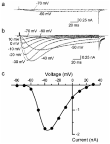
          Sensory neurons in the dorsal root ganglion express two kinds of tetrodotoxin resistant (TTX-R) isoforms of voltage-gated sodium channels, Na V1.8 and Na V1.9. These isoforms play key roles in the pathophysiology of chronic pain. Of special interest is Na V1.9: our previous studies revealed a unique property of the Na V1.9 current, i.e., the Na V1.9 current shows a gradual and notable up-regulation of the peak amplitude during recording (“spontaneous augmentation of Na V1.9”). However, the mechanism underlying the spontaneous augmentation of Na V1.9 is still unclear. In this study, we examined the effects of protein kinases A and C (PKA and PKC), on the spontaneous augmentation of Na V1.9. The spontaneous augmentation of the Na V1.9 current was significantly suppressed by activation of PKA, whereas activation of PKA did not affect the voltage dependence of inactivation for the Na V1.9 current. On the contrary, the finding that activation of PKC can affect the voltage dependence of inactivation for Na V1.9 in the perforated patch recordings, where the augmentation does not occur, suggests that the effects of PMA are independent of the augmentation process. These results indicate that the spontaneous augmentation of Na V1.9 was regulated directly by PKA, and indirectly by PKC.

          Related collections

          Most cited references39

          • Record: found
          • Abstract: found
          • Article: not found

          Improved patch-clamp techniques for high-resolution current recording from cells and cell-free membrane patches.

          1. The extracellular patch clamp method, which first allowed the detection of single channel currents in biological membranes, has been further refined to enable higher current resolution, direct membrane patch potential control, and physical isolation of membrane patches. 2. A description of a convenient method for the fabrication of patch recording pipettes is given together with procedures followed to achieve giga-seals i.e. pipette-membrane seals with resistances of 10(9) - 10(11) omega. 3. The basic patch clamp recording circuit, and designs for improved frequency response are described along with the present limitations in recording the currents from single channels. 4. Procedures for preparation and recording from three representative cell types are given. Some properties of single acetylcholine-activated channels in muscle membrane are described to illustrate the improved current and time resolution achieved with giga-seals. 5. A description is given of the various ways that patches of membrane can be physically isolated from cells. This isolation enables the recording of single channel currents with well-defined solutions on both sides of the membrane. Two types of isolated cell-free patch configurations can be formed: an inside-out patch with its cytoplasmic membrane face exposed to the bath solution, and an outside-out patch with its extracellular membrane face exposed to the bath solution. 6. The application of the method for the recording of ionic currents and internal dialysis of small cells is considered. Single channel resolution can be achieved when recording from whole cells, if the cell diameter is small (less than 20 micrometer). 7. The wide range of cell types amenable to giga-seal formation is discussed.
            Bookmark
            • Record: found
            • Abstract: found
            • Article: not found

            The tetrodotoxin-resistant sodium channel SNS has a specialized function in pain pathways.

            Many damage-sensing neurons express tetrodotoxin (TTX)-resistant voltage-gated sodium channels. Here we examined the role of the sensory-neuron-specific (SNS) TTX-resistant sodium channel alpha subunit in nociception and pain by constructing sns-null mutant mice. These mice expressed only TTX-sensitive sodium currents on step depolarizations from normal resting potentials, showing that all slow TTX-resistant currents are encoded by the sns gene. Null mutants were viable, fertile and apparently normal, although lowered thresholds of electrical activation of C-fibers and increased current densities of TTX-sensitive channels demonstrated compensatory upregulation of TTX-sensitive currents in sensory neurons. Behavioral studies demonstrated a pronounced analgesia to noxious mechanical stimuli, small deficits in noxious thermoreception and delayed development of inflammatory hyperalgesia. These data show that SNS is involved in pain pathways and suggest that blockade of SNS expression or function may produce analgesia without side effects.
              Bookmark
              • Record: found
              • Abstract: found
              • Article: not found

              Contribution of Na(v)1.8 sodium channels to action potential electrogenesis in DRG neurons.

              C-type dorsal root ganglion (DRG) neurons can generate tetrodotoxin-resistant (TTX-R) sodium-dependent action potentials. However, multiple sodium channels are expressed in these neurons, and the molecular identity of the TTX-R sodium channels that contribute to action potential production in these neurons has not been established. In this study, we used current-clamp recordings to compare action potential electrogenesis in Na(v)1.8 (+/+) and (-/-) small DRG neurons maintained for 2-8 h in vitro to examine the role of sodium channel Na(v)1.8 (alpha-SNS) in action potential electrogenesis. Although there was no significant difference in resting membrane potential, input resistance, current threshold, or voltage threshold in Na(v)1.8 (+/+) and (-/-) DRG neurons, there were significant differences in action potential electrogenesis. Most Na(v)1.8 (+/+) neurons generate all-or-none action potentials, whereas most of Na(v)1.8 (-/-) neurons produce smaller graded responses. The peak of the response was significantly reduced in Na(v)1.8 (-/-) neurons [31.5 +/- 2.2 (SE) mV] compared with Na(v)1.8 (+/+) neurons (55.0 +/- 4.3 mV). The maximum rise slope was 84.7 +/- 11.2 mV/ms in Na(v)1.8 (+/+) neurons, significantly faster than in Na(v)1.8 (-/-) neurons where it was 47.2 +/- 1.3 mV/ms. Calculations based on the action potential overshoot in Na(v)1.8 (+/+) and (-/-) neurons, following blockade of Ca(2+) currents, indicate that Na(v)1.8 contributes a substantial fraction (80-90%) of the inward membrane current that flows during the rising phase of the action potential. We found that fast TTX-sensitive Na(+) channels can produce all-or-none action potentials in some Na(v)1.8 (-/-) neurons but, presumably as a result of steady-state inactivation of these channels, electrogenesis in Na(v)1.8 (-/-) neurons is more sensitive to membrane depolarization than in Na(v)1.8 (+/+) neurons, and, in the absence of Na(v)1.8, is attenuated with even modest depolarization. These observations indicate that Na(v)1.8 contributes substantially to action potential electrogenesis in C-type DRG neurons.
                Bookmark

                Author and article information

                Journal
                Mar Drugs
                MD
                Marine Drugs
                Molecular Diversity Preservation International
                1660-3397
                2010
                19 March 2010
                : 8
                : 3
                : 728-740
                Affiliations
                [1 ] Technical Center, Hiroshima University, Hiroshima, 734-8551, Japan
                [2 ] Department of Neurophysiology, Graduate School of Biomedical Sciences, Hiroshima University, Hiroshima, 734-8551, Japan; E-Mails: tei0322@ 123456hiroshima-u.ac.jp (T.Z.); uryu@ 123456asahi.ac.jp (N.U.); ogatan@ 123456hiroshima-u.ac.jp (N.O.)
                Author notes
                * Author to whom correspondence should be addressed; E-Mail: kakimura@ 123456hiroshima-u.ac.jp ; Tel.: +81-82-257-5127; Fax: +81-82-257-5127.
                Article
                marinedrugs-08-00728
                10.3390/md8030728
                2857352
                20411123
                124c2c5c-b606-4bb8-8618-f6e3078e0ee9
                © 2010 by the authors; licensee Molecular Diversity Preservation International, Basel, Switzerland

                This article is an open-access article distributed under the terms and conditions of the Creative Commons Attribution license ( http://creativecommons.org/licenses/by/3.0/).

                History
                : 15 January 2010
                : 8 February 2010
                : 18 March 2010
                Categories
                Article

                Pharmacology & Pharmaceutical medicine
                patch clamp,pkc,dorsal root ganglion,tetrodotoxin,pka,na+ channel

                Comments

                Comment on this article