0
views
0
recommends
+1 Recommend
0 collections
    0
    shares
      • Record: found
      • Abstract: found
      • Article: found
      Is Open Access

      Highly Multiplexed Single-Cell In Situ RNA and DNA Analysis by Consecutive Hybridization

      research-article

      Read this article at

      Bookmark
          There is no author summary for this article yet. Authors can add summaries to their articles on ScienceOpen to make them more accessible to a non-specialist audience.

          Abstract

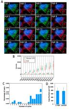
          The ability to comprehensively profile nucleic acids in individual cells in their natural spatial contexts is essential to advance our understanding of biology and medicine. Here, we report a novel method for spatial transcriptomics and genomics analysis. In this method, every nucleic acid molecule is detected as a fluorescent spot at its natural cellular location throughout the cycles of consecutive fluorescence in situ hybridization (C-FISH). In each C-FISH cycle, fluorescent oligonucleotide probes hybridize to the probes applied in the previous cycle, and also introduce the binding sites for the next cycle probes. With reiterative cycles of hybridization, imaging and photobleaching, the identities of the varied nucleic acids are determined by their unique color sequences. To demonstrate the feasibility of this method, we show that transcripts or genomic loci in single cells can be unambiguously quantified with 2 fluorophores and 16 C-FISH cycles or with 3 fluorophores and 9 C-FISH cycles. Without any error correction, the error rates obtained using the raw data are close to zero. These results indicate that C-FISH potentially enables tens of thousands (2 16 = 65,536 or 3 9 = 19,683) of different transcripts or genomic loci to be precisely profiled in individual cells in situ.

          Related collections

          Most cited references39

          • Record: found
          • Abstract: found
          • Article: not found

          Proteomics. Tissue-based map of the human proteome.

          Resolving the molecular details of proteome variation in the different tissues and organs of the human body will greatly increase our knowledge of human biology and disease. Here, we present a map of the human tissue proteome based on an integrated omics approach that involves quantitative transcriptomics at the tissue and organ level, combined with tissue microarray-based immunohistochemistry, to achieve spatial localization of proteins down to the single-cell level. Our tissue-based analysis detected more than 90% of the putative protein-coding genes. We used this approach to explore the human secretome, the membrane proteome, the druggable proteome, the cancer proteome, and the metabolic functions in 32 different tissues and organs. All the data are integrated in an interactive Web-based database that allows exploration of individual proteins, as well as navigation of global expression patterns, in all major tissues and organs in the human body. Copyright © 2015, American Association for the Advancement of Science.
            Bookmark
            • Record: found
            • Abstract: found
            • Article: not found

            RNA imaging. Spatially resolved, highly multiplexed RNA profiling in single cells.

            Knowledge of the expression profile and spatial landscape of the transcriptome in individual cells is essential for understanding the rich repertoire of cellular behaviors. Here, we report multiplexed error-robust fluorescence in situ hybridization (MERFISH), a single-molecule imaging approach that allows the copy numbers and spatial localizations of thousands of RNA species to be determined in single cells. Using error-robust encoding schemes to combat single-molecule labeling and detection errors, we demonstrated the imaging of 100 to 1000 distinct RNA species in hundreds of individual cells. Correlation analysis of the ~10(4) to 10(6) pairs of genes allowed us to constrain gene regulatory networks, predict novel functions for many unannotated genes, and identify distinct spatial distribution patterns of RNAs that correlate with properties of the encoded proteins.
              Bookmark
              • Record: found
              • Abstract: found
              • Article: not found

              Transcriptome-scale super-resolved imaging in tissues by RNA seqFISH+

              Imaging the transcriptome in situ with high accuracy has been a major challenge in single cell biology, particularly hindered by the limits of optical resolution and the density of transcripts in single cells 1–5 . Here, we demonstrate seqFISH+, that can image the mRNAs for 10,000 genes in single cells with high accuracy and sub-diffraction-limit resolution, in the mouse brain cortex, subventricular zone, and the olfactory bulb, using a standard confocal microscope. The transcriptome level profiling of seqFISH+ allows unbiased identification of cell classes and their spatial organization in tissues. In addition, seqFISH+ reveals subcellular mRNA localization patterns in cells and ligand-receptor pairs across neighboring cells. This technology demonstrates the ability to generate spatial cell atlases and to perform discovery-driven studies of biological processes in situ.
                Bookmark

                Author and article information

                Contributors
                Role: Academic Editor
                Role: Academic Editor
                Journal
                Molecules
                Molecules
                molecules
                Molecules
                MDPI
                1420-3049
                23 October 2020
                November 2020
                : 25
                : 21
                : 4900
                Affiliations
                Biodesign Institute & School of Molecular Sciences, Arizona State University, Tempe, AZ 85287, USA; lxiao15@ 123456asu.edu (L.X.); Renjie.Liao@ 123456asu.edu (R.L.)
                Author notes
                [* ]Correspondence: jiaguo@ 123456asu.edu ; Tel.: +1-480-727-2096
                Author information
                https://orcid.org/0000-0001-8036-5349
                Article
                molecules-25-04900
                10.3390/molecules25214900
                7660199
                33113917
                12fc521d-a1f8-4d5d-8246-1d875a2021ff
                © 2020 by the authors.

                Licensee MDPI, Basel, Switzerland. This article is an open access article distributed under the terms and conditions of the Creative Commons Attribution (CC BY) license ( http://creativecommons.org/licenses/by/4.0/).

                History
                : 27 September 2020
                : 22 October 2020
                Categories
                Article

                fluorescence in situ hybridization,fish,transcripts,genomic loci,transcriptomics,genomics

                Comments

                Comment on this article