25
views
0
recommends
+1 Recommend
1 collections
    0
    shares
      • Record: found
      • Abstract: found
      • Article: found
      Is Open Access

      Management of glucocorticoids-induced osteoporosis: role of teriparatide

      review-article

      Read this article at

      Bookmark
          There is no author summary for this article yet. Authors can add summaries to their articles on ScienceOpen to make them more accessible to a non-specialist audience.

          Abstract

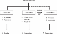
          Glucocorticoids (GC)-induced osteoporosis (GIOP) is the most common cause of secondary osteoporosis, which leads to an increased fracture risk in patients. The normal bone turnover depends on a balance between osteoblasts and osteoclasts activity and GC can cause a rapid bone loss, decreasing bone formation and increasing bone resorption. The decreased bone formation is mainly due to the GC-induced apoptosis of both osteoblasts and osteocytes, while the increased bone resorption is due to the increased life-span of pre-existing osteoclasts. Bisphosphonates are clearly effective in preventing and treating GIOP but anabolic therapeutic strategies are the new promising therapeutic alternative. Experimental and clinical studies indicate that teriparatide, the active (1–34) parathyroid hormone (PTH) molecule, is efficacious for the treatment of GIOP, being able to induce an increase in bone mass in these patients. Intermittent administration of human PTH (1–34) stimulates bone formation by increasing osteoblast number. Additionally, human PTH (1–34) modulates the level and/or activity of locally produced growth factors and cytokines. Teriparatide has been demonstrated in several clinical studies to significantly decrease the incidence of fractures in patients affected by GIOP. It has recently received an indication for GIOP and its label indication has also been expanded.

          Most cited references80

          • Record: found
          • Abstract: found
          • Article: not found

          Osteoporosis prevention, diagnosis, and therapy.

          (2001)
          To clarify the factors associated with prevention, diagnosis, and treatment of osteoporosis, and to present the most recent information available in these areas. From March 27-29, 2000, a nonfederal, nonadvocate, 13-member panel was convened, representing the fields of internal medicine, family and community medicine, endocrinology, epidemiology, orthopedic surgery, gerontology, rheumatology, obstetrics and gynecology, preventive medicine, and cell biology. Thirty-two experts from these fields presented data to the panel and an audience of 699. Primary sponsors were the National Institute of Arthritis and Musculoskeletal and Skin Diseases and the National Institutes of Health Office of Medical Applications of Research. MEDLINE was searched for January 1995 through December 1999, and a bibliography of 2449 references provided to the panel. Experts prepared abstracts for presentations with relevant literature citations. Scientific evidence was given precedence over anecdotal experience. The panel, answering predefined questions, developed conclusions based on evidence presented in open forum and the literature. The panel composed a draft statement, which was read and circulated to the experts and the audience for public discussion. The panel resolved conflicts and released a revised statement at the end of the conference. The draft statement was posted on the Web on March 30, 2000, and updated with the panel's final revisions within a few weeks. Though prevalent in white postmenopausal women, osteoporosis occurs in all populations and at all ages and has significant physical, psychosocial, and financial consequences. Risks for osteoporosis (reflected by low bone mineral density [BMD]) and for fracture overlap but are not identical. More attention should be paid to skeletal health in persons with conditions associated with secondary osteoporosis. Clinical risk factors have an important but poorly validated role in determining who should have BMD measurement, in assessing fracture risk, and in determining who should be treated. Adequate calcium and vitamin D intake is crucial to develop optimal peak bone mass and to preserve bone mass throughout life. Supplementation with these 2 nutrients may be necessary in persons not achieving recommended dietary intake. Gonadal steroids are important determinants of peak and lifetime bone mass in men, women, and children. Regular exercise, especially resistance and high-impact activities, contributes to development of high peak bone mass and may reduce risk of falls in older persons. Assessment of bone mass, identification of fracture risk, and determination of who should be treated are the optimal goals when evaluating patients for osteoporosis. Fracture prevention is the primary treatment goal for patients with osteoporosis. Several treatments have been shown to reduce the risk of osteoporotic fractures, including those that enhance bone mass and reduce the risk or consequences of falls. Adults with vertebral, rib, hip, or distal forearm fractures should be evaluated for osteoporosis and given appropriate therapy.
            Bookmark
            • Record: found
            • Abstract: found
            • Article: not found

            Teriparatide or alendronate in glucocorticoid-induced osteoporosis.

            Bisphosphonate therapy is the current standard of care for the prevention and treatment of glucocorticoid-induced osteoporosis. Studies of anabolic therapy in patients who are receiving long-term glucocorticoids and are at high risk for fracture are lacking. In an 18-month randomized, double-blind, controlled trial, we compared teriparatide with alendronate in 428 women and men with osteoporosis (ages, 22 to 89 years) who had received glucocorticoids for at least 3 months (prednisone equivalent, 5 mg daily or more). A total of 214 patients received 20 microg of teriparatide once daily, and 214 received 10 mg of alendronate once daily. The primary outcome was the change in bone mineral density at the lumbar spine. Secondary outcomes included changes in bone mineral density at the total hip and in markers of bone turnover, the time to changes in bone mineral density, the incidence of fractures, and safety. At the last measurement, the mean (+/-SE) bone mineral density at the lumbar spine had increased more in the teriparatide group than in the alendronate group (7.2+/-0.7% vs. 3.4+/-0.7%, P<0.001). A significant difference between the groups was reached by 6 months (P<0.001). At 12 months, bone mineral density at the total hip had increased more in the teriparatide group. Fewer new vertebral fractures occurred in the teriparatide group than in the alendronate group (0.6% vs. 6.1%, P=0.004); the incidence of nonvertebral fractures was similar in the two groups (5.6% vs. 3.7%, P=0.36). Significantly more patients in the teriparatide group had at least one elevated measure of serum calcium. Among patients with osteoporosis who were at high risk for fracture, bone mineral density increased more in patients receiving teriparatide than in those receiving alendronate. (ClinicalTrials.gov number, NCT00051558 [ClinicalTrials.gov].). Copyright 2007 Massachusetts Medical Society.
              Bookmark
              • Record: found
              • Abstract: found
              • Article: not found

              Glucocorticoids act directly on osteoblasts and osteocytes to induce their apoptosis and reduce bone formation and strength.

              Whether the negative impact of excess glucocorticoids on the skeleton is due to direct effects on bone cells, indirect effects on extraskeletal tissues, or both is unknown. To determine the contribution of direct effects of glucocorticoids on osteoblastic/osteocytic cells in vivo, we blocked glucocorticoid action on these cells via transgenic expression of 11beta-hydroxysteroid dehydrogenase type 2, an enzyme that inactivates glucocorticoids. Osteoblast/osteocyte-specific expression was achieved by insertion of the 11beta-hydroxysteroid dehydrogenase type 2 cDNA downstream from the osteoblast-specific osteocalcin promoter. The transgene did not affect normal bone development or turnover as demonstrated by identical bone density, strength, and histomorphometry in adult transgenic and wild-type animals. Administration of excess glucocorticoids induced equivalent bone loss in wild-type and transgenic mice. As expected, cancellous osteoclasts were unaffected by the transgene. However, the increase in osteoblast apoptosis that occurred in wild-type mice was prevented in transgenic mice. Consistent with this, osteoblasts, osteoid area, and bone formation rate were significantly higher in glucocorticoid-treated transgenic mice compared with glucocorticoid-treated wild-type mice. Glucocorticoid-induced osteocyte apoptosis was also prevented in transgenic mice. Strikingly, the loss of vertebral compression strength observed in glucocorticoid-treated wild-type mice was prevented in the transgenic mice, despite equivalent bone loss. These results demonstrate for the first time that excess glucocorticoids directly affect bone forming cells in vivo. Furthermore, our results suggest that glucocorticoid-induced loss of bone strength results in part from increased death of osteocytes, independent of bone loss.
                Bookmark

                Author and article information

                Journal
                Ther Clin Risk Manag
                Therapeutics and Clinical Risk Management
                Therapeutics and Clinical Risk Management
                Dove Medical Press
                1176-6336
                1178-203X
                2009
                2009
                4 May 2009
                : 5
                : 305-310
                Affiliations
                [1 ]Dipartimento di Fisiopatologia Medica, Policlinico Umberto I, Università degli Studi Sapienza di Roma, Italy;
                [2 ]Dipartimento di Medicina Interna, Policlinico S Orsola Malpighi, Bologna, Italy
                Author notes
                Correspondence: Silvia Migliaccio, Dipartimento di Fisiopatologia Medica, Università Sapienza di Roma, Italy, Tel +39 06 4997 0721, Fax + 39 06 446 1450, Email silvia.migliaccio@ 123456uniroma1.it
                Article
                tcrm-5-305
                2697534
                19536312
                1305a666-442f-4bb8-80ae-3beef7986a1c
                © 2009 Migliaccio et al, publisher and licensee Dove Medical Press Ltd.

                This is an Open Access article which permits unrestricted noncommercial use, provided the original work is properly cited.

                History
                Categories
                Review

                Medicine
                teriparatide,osteoporosis,osteoclasts,osteoblasts,glucocorticoids
                Medicine
                teriparatide, osteoporosis, osteoclasts, osteoblasts, glucocorticoids

                Comments

                Comment on this article