16
views
0
recommends
+1 Recommend
0 collections
    0
    shares
      • Record: found
      • Abstract: found
      • Article: found
      Is Open Access

      Enpp1 is an anti-aging factor that regulates Klotho under phosphate overload conditions

      research-article

      Read this article at

      Bookmark
          There is no author summary for this article yet. Authors can add summaries to their articles on ScienceOpen to make them more accessible to a non-specialist audience.

          Abstract

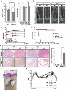
          Control of phosphate metabolism is crucial to regulate aging in mammals. Klotho is a well-known anti-aging factor that regulates phosphate metabolism: mice mutant or deficient in Klotho exhibit phenotypes resembling human aging. Here we show that ectonucleotide pyrophosphatase/phosphodiesterase 1 (Enpp1) is required for Klotho expression under phosphate overload conditions. Loss-of-function Enpp1 ttw/ttw mice under phosphate overload conditions exhibited phenotypes resembling human aging and Klotho mutants, such as short life span, arteriosclerosis and osteoporosis, with elevated serum 1,25(OH) 2D 3 levels. Enpp1 ttw/ttw mice also exhibited significantly reduced renal Klotho expression under phosphate overload conditions, and aging phenotypes in these mice were rescued by Klotho overexpression, a low vitamin D diet or vitamin D receptor knockout. These findings indicate that Enpp1 plays a crucial role in regulating aging via Klotho expression under phosphate overload conditions.

          Related collections

          Most cited references54

          • Record: found
          • Abstract: found
          • Article: not found

          Klotho deficiency causes vascular calcification in chronic kidney disease.

          Soft-tissue calcification is a prominent feature in both chronic kidney disease (CKD) and experimental Klotho deficiency, but whether Klotho deficiency is responsible for the calcification in CKD is unknown. Here, wild-type mice with CKD had very low renal, plasma, and urinary levels of Klotho. In humans, we observed a graded reduction in urinary Klotho starting at an early stage of CKD and progressing with loss of renal function. Despite induction of CKD, transgenic mice that overexpressed Klotho had preserved levels of Klotho, enhanced phosphaturia, better renal function, and much less calcification compared with wild-type mice with CKD. Conversely, Klotho-haploinsufficient mice with CKD had undetectable levels of Klotho, worse renal function, and severe calcification. The beneficial effect of Klotho on vascular calcification was a result of more than its effect on renal function and phosphatemia, suggesting a direct effect of Klotho on the vasculature. In vitro, Klotho suppressed Na(+)-dependent uptake of phosphate and mineralization induced by high phosphate and preserved differentiation in vascular smooth muscle cells. In summary, Klotho is an early biomarker for CKD, and Klotho deficiency contributes to soft-tissue calcification in CKD. Klotho ameliorates vascular calcification by enhancing phosphaturia, preserving glomerular filtration, and directly inhibiting phosphate uptake by vascular smooth muscle. Replacement of Klotho may have therapeutic potential for CKD.
            Bookmark
            • Record: found
            • Abstract: found
            • Article: not found

            Autosomal dominant hypophosphataemic rickets is associated with mutations in FGF23.

            (2000)
            Proper serum phosphate concentrations are maintained by a complex and poorly understood process. Identification of genes responsible for inherited disorders involving disturbances in phosphate homeostasis may provide insight into the pathways that regulate phosphate balance. Several hereditary disorders of isolated phosphate wasting have been described, including X-linked hypophosphataemic rickets (XLH), hypophosphataemic bone disease (HBD), hereditary hypophosphataemic rickets with hypercalciuria (HHRH) and autosomal dominant hypophosphataemic rickets (ADHR). Inactivating mutations of the gene PHEX, encoding a member of the neutral endopeptidase family of proteins, are responsible for XLH (refs 6,7). ADHR (MIM 193100) is characterized by low serum phosphorus concentrations, rickets, osteomalacia, lower extremity deformities, short stature, bone pain and dental abscesses. Here we describe a positional cloning approach used to identify the ADHR gene which included the annotation of 37 genes within 4 Mb of genomic sequence. We identified missense mutations in a gene encoding a new member of the fibroblast growth factor (FGF) family, FGF23. These mutations in patients with ADHR represent the first mutations found in a human FGF gene.
              Bookmark
              • Record: found
              • Abstract: found
              • Article: not found

              Cloning and characterization of FGF23 as a causative factor of tumor-induced osteomalacia.

              Tumor-induced osteomalacia (TIO) is one of the paraneoplastic diseases characterized by hypophosphatemia caused by renal phosphate wasting. Because removal of responsible tumors normalizes phosphate metabolism, an unidentified humoral phosphaturic factor is believed to be responsible for this syndrome. To identify the causative factor of TIO, we obtained cDNA clones that were abundantly expressed only in a tumor causing TIO and constructed tumor-specific cDNA contigs. Based on the sequence of one major contig, we cloned 2,270-bp cDNA, which turned out to encode fibroblast growth factor 23 (FGF23). Administration of recombinant FGF23 decreased serum phosphate in mice within 12 h. When Chinese hamster ovary cells stably expressing FGF23 were s.c. implanted into nude mice, hypophosphatemia with increased renal phosphate clearance was observed. In addition, a high level of serum alkaline phosphatase, low 1,25-dihydroxyvitamin D, deformity of bone, and impairment of body weight gain became evident. Histological examination showed marked increase of osteoid and widening of growth plate. Thus, continuous production of FGF23 reproduced clinical, biochemical, and histological features of TIO in vivo. Analyses for recombinant FGF23 products produced by Chinese hamster ovary cells indicated proteolytic cleavage of FGF23 at the RXXR motif. Recent genetic study indicates that missense mutations in this RXXR motif of FGF23 are responsible for autosomal dominant hypophosphatemic rickets, another hypophosphatemic disease with similar features to TIO. We conclude that overproduction of FGF23 causes TIO, whereas mutations in the FGF23 gene result in autosomal dominant hypophosphatemic rickets possibly by preventing proteolytic cleavage and enhancing biological activity of FGF23.
                Bookmark

                Author and article information

                Contributors
                miyamoto@z5.keio.jp
                Journal
                Sci Rep
                Sci Rep
                Scientific Reports
                Nature Publishing Group UK (London )
                2045-2322
                10 August 2017
                10 August 2017
                2017
                : 7
                : 7786
                Affiliations
                [1 ]ISNI 0000 0004 1936 9959, GRID grid.26091.3c, Department of Orthopedic Surgery, , Keio University School of Medicine, ; 35 Shinano-machi, Shinjuku-ku, Tokyo, 160-8582 Japan
                [2 ]ISNI 0000 0004 1936 9959, GRID grid.26091.3c, Department of Advanced Therapy for Musculoskeletal Disorders, , Keio University School of Medicine, ; 35 Shinano-machi, Shinjuku-ku, Tokyo, 160-8582 Japan
                [3 ]ISNI 0000 0004 1936 9959, GRID grid.26091.3c, Department of Musculoskeletal Reconstruction and Regeneration Surgery, , Keio University School of Medicine, ; 35 Shinano-machi, Shinjuku-ku, Tokyo, 160-8582 Japan
                [4 ]ISNI 0000 0004 1936 9959, GRID grid.26091.3c, Division of Oral and Maxillofacial Surgery, Department of Dentistry and Oral Surgery, , Keio University School of Medicine, ; 35 Shinano-machi, Shinjuku-ku, Tokyo, 160-8582 Japan
                [5 ]ISNI 0000000123090000, GRID grid.410804.9, Center for Molecular Medicine, , Jichi Medical University, ; Shimotsuke, Tochigi, 329-0498 Japan
                [6 ]ISNI 0000 0004 0377 2137, GRID grid.416629.e, Department of Bone and Mineral Research, , Osaka Medical Center and Research Institute for Maternal and Child Health, ; Izumi, Osaka, 594-1101 Japan
                [7 ]ISNI 0000 0001 1092 3579, GRID grid.267335.6, Fujii Memorial Institute of Medical Sciences, , Tokushima University, Tokushima, ; Tokushima, 770-8503 Japan
                Author information
                http://orcid.org/0000-0003-3610-3469
                Article
                7341
                10.1038/s41598-017-07341-2
                5552841
                28798354
                13b19d2f-5828-45b7-8cd2-e4e67bd67bab
                © The Author(s) 2017

                Open Access This article is licensed under a Creative Commons Attribution 4.0 International License, which permits use, sharing, adaptation, distribution and reproduction in any medium or format, as long as you give appropriate credit to the original author(s) and the source, provide a link to the Creative Commons license, and indicate if changes were made. The images or other third party material in this article are included in the article’s Creative Commons license, unless indicated otherwise in a credit line to the material. If material is not included in the article’s Creative Commons license and your intended use is not permitted by statutory regulation or exceeds the permitted use, you will need to obtain permission directly from the copyright holder. To view a copy of this license, visit http://creativecommons.org/licenses/by/4.0/.

                History
                : 16 February 2017
                : 6 June 2017
                Categories
                Article
                Custom metadata
                © The Author(s) 2017

                Uncategorized
                Uncategorized

                Comments

                Comment on this article