8
views
0
recommends
+1 Recommend
0 collections
    0
    shares
      • Record: found
      • Abstract: found
      • Article: found
      Is Open Access

      MicroRNA‐382 silencing induces a mitonuclear protein imbalance and activates the mitochondrial unfolded protein response in muscle cells

      research-article

      Read this article at

      Bookmark
          There is no author summary for this article yet. Authors can add summaries to their articles on ScienceOpen to make them more accessible to a non-specialist audience.

          Abstract

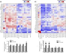
          Proper mitochondrial function plays a central role in cellular metabolism. Various diseases as well as aging are associated with diminished mitochondrial function. Previously, we identified 19 miRNAs putatively involved in the regulation of mitochondrial metabolism in skeletal muscle, a highly metabolically active tissue. In the current study, these 19 miRNAs were individually silenced in C2C12 myotubes using antisense oligonucleotides, followed by measurement of the expression of 27 genes known to play a major role in regulating mitochondrial metabolism. Based on the outcomes, we then focused on miR‐382‐5p and identified pathways affected by its silencing using microarrays, investigated protein expression, and studied cellular respiration. Silencing of miRNA‐382‐5p significantly increased the expression of several genes involved in mitochondrial dynamics and biogenesis. Conventional microarray analysis in C2C12 myotubes silenced for miRNA‐382‐5p revealed a collective downregulation of mitochondrial ribosomal proteins and respiratory chain proteins. This effect was accompanied by an imbalance between mitochondrial proteins encoded by the nuclear and mitochondrial DNA (1.35‐fold, p < 0.01) and an induction of HSP60 protein (1.31‐fold, p < 0.05), indicating activation of the mitochondrial unfolded protein response (mtUPR). Furthermore, silencing of miR‐382‐5p reduced basal oxygen consumption rate by 14% ( p < 0.05) without affecting mitochondrial content, pointing towards a more efficient mitochondrial function as a result of improved mitochondrial quality control. Taken together, silencing of miR‐382‐5p induces a mitonuclear protein imbalance and activates the mtUPR in skeletal muscle, a phenomenon that was previously associated with improved longevity.

          Related collections

          Most cited references20

          • Record: found
          • Abstract: found
          • Article: not found

          Adipose mitochondrial biogenesis is suppressed in db/db and high-fat diet-fed mice and improved by rosiglitazone.

          The objective of this study was to further establish and confirm the relationship of adipose mitochondrial biogenesis in diabetes/obesity and the effects of rosiglitazone (RSG), a peroxisome proliferator-activated receptor (PPAR) gamma agonist, by systematically analyzing mitochondrial gene expression and function in two mouse models of obesity and type 2 diabetes. Using microarray technology, adipose mitochondrial gene transcription was studied in db/db, high-fat diet-fed C57BL/6 (HFD) and respective control mice with or without RSG treatment. The findings were extended using mitochondrial staining, DNA quantification, and measurements of citrate synthase activity. In db/db and HFD mice, gene transcripts associated with mitochondrial ATP production, energy uncoupling, mitochondrial ribosomal proteins, outer and inner membrane translocases, and mitochondrial heat-shock proteins were decreased in abundance, compared with db/+ and standard-fat diet-fed control mice, respectively. RSG dose-dependently increased these transcripts in both db/db and HFD mice and induced transcription of mitochondrial structural proteins and cellular antioxidant enzymes responsible for removal of reactive oxygen species generated by increased mitochondrial activity. Transcription factors, including PPAR coactivator (PGC)-1beta, PGC-1alpha, estrogen-related receptor alpha, and PPARalpha, were suppressed in both models and induced by RSG. The effects of RSG on adipose mitochondrial genes were confirmed by quantitative RT-PCR and further supported by mitochondrial staining, mitochondrial DNA quantification, and citrate synthase activity. Adipose mitochondrial biogenesis was overwhelmingly suppressed in both mouse models of diabetes/obesity and globally induced by RSG. These findings suggest an important role of adipose mitochondria in diabetes/obesity and the potential for new treatment approaches targeting adipose mitochondria.
            Bookmark
            • Record: found
            • Abstract: found
            • Article: not found

            Multilayered genetic and omics dissection of mitochondrial activity in a mouse reference population.

            The manner by which genotype and environment affect complex phenotypes is one of the fundamental questions in biology. In this study, we quantified the transcriptome--a subset of the metabolome--and, using targeted proteomics, quantified a subset of the liver proteome from 40 strains of the BXD mouse genetic reference population on two diverse diets. We discovered dozens of transcript, protein, and metabolite QTLs, several of which linked to metabolic phenotypes. Most prominently, Dhtkd1 was identified as a primary regulator of 2-aminoadipate, explaining variance in fasted glucose and diabetes status in both mice and humans. These integrated molecular profiles also allowed further characterization of complex pathways, particularly the mitochondrial unfolded protein response (UPR(mt)). UPR(mt) shows strikingly variant responses at the transcript and protein level that are remarkably conserved among C. elegans, mice, and humans. Overall, these examples demonstrate the value of an integrated multilayered omics approach to characterize complex metabolic phenotypes. Copyright © 2014 Elsevier Inc. All rights reserved.
              Bookmark
              • Record: found
              • Abstract: found
              • Article: not found

              Intramuscular heat shock protein 72 and heme oxygenase-1 mRNA are reduced in patients with type 2 diabetes: evidence that insulin resistance is associated with a disturbed antioxidant defense mechanism.

              To examine whether genes associated with cellular defense against oxidative stress are associated with insulin sensitivity, patients with type 2 diabetes (n = 7) and age-matched (n = 5) and young (n = 9) control subjects underwent a euglycemic-hyperinsulinemic clamp for 120 min. Muscle samples were obtained before and after the clamp and analyzed for heat shock protein (HSP)72 and heme oxygenase (HO)-1 mRNA, intramuscular triglyceride content, and the maximal activities of beta-hydroxyacyl-CoA dehydrogenase (beta-HAD) and citrate synthase (CS). Basal expression of both HSP72 and HO-1 mRNA were lower (P < 0.05) by 33 and 55%, respectively, when comparing diabetic patients with age-matched and young control subjects, with no differences between the latter groups. Both basal HSP72 (r = 0.75, P < 0.001) and HO-1 (r = 0.50, P < 0.05) mRNA expression correlated with the glucose infusion rate during the clamp. Significant correlations were also observed between HSP72 mRNA and both beta-HAD (r = 0.61, P < 0.01) and CS (r = 0.65, P < 0.01). HSP72 mRNA was induced (P < 0.05) by the clamp in all groups. Although HO-1 mRNA was unaffected by the clamp in both the young and age-matched control subjects, it was increased (P < 0.05) approximately 70-fold in the diabetic patients after the clamp. These data demonstrate that genes involved in providing cellular protection against oxidative stress are defective in patients with type 2 diabetes and correlate with insulin-stimulated glucose disposal and markers of muscle oxidative capacity. The data provide new evidence that the pathogenesis of type 2 diabetes involves perturbations to the antioxidant defense mechanism within skeletal muscle.
                Bookmark

                Author and article information

                Contributors
                j.hoeks@maastrichtuniversity.nl
                Journal
                J Cell Physiol
                J. Cell. Physiol
                10.1002/(ISSN)1097-4652
                JCP
                Journal of Cellular Physiology
                John Wiley and Sons Inc. (Hoboken )
                0021-9541
                1097-4652
                11 November 2018
                May 2019
                : 234
                : 5 ( doiID: 10.1002/jcp.v234.5 )
                : 6601-6610
                Affiliations
                [ 1 ] Department of Nutrition and Movement Sciences NUTRIM School of Nutrition and Translational Research in Metabolism, Maastricht University The Netherlands
                [ 2 ] Laboratory of Integrative Systems Physiology, Institute of Bioengineering, École Polytechnique Fédérale de Lausanne Lausanne Switzerland
                [ 3 ] Division of Human Nutrition Nutrition, Metabolism and Genomics Group, Wageningen University Wageningen The Netherlands
                Author notes
                [*] [* ] Correspondence Joris Hoeks, Department of Nutrition and Movement Sciences, NUTRIM School of Nutrition and Translational Research in Metabolism, Maastricht University, PO Box 616, 6200MD Maastricht, The Netherlands. Email: j.hoeks@ 123456maastrichtuniversity.nl

                Author information
                http://orcid.org/0000-0002-0265-0870
                Article
                JCP27401
                10.1002/jcp.27401
                6344277
                30417335
                140f4bf7-9921-4765-9511-0da48a7b9f8b
                © 2018 The Authors. Journal of Cellular Physiology Published by Wiley Periodicals, Inc.

                This is an open access article under the terms of the http://creativecommons.org/licenses/by-n/4.0/ License, which permits use, distribution and reproduction in any medium, provided the original work is properly cited and is not used for commercial purposes.

                History
                : 22 November 2017
                : 17 August 2018
                Page count
                Figures: 5, Tables: 1, Pages: 10, Words: 6364
                Funding
                Funded by: Systems X
                Award ID: SySX.ch 2013/153
                Funded by: Velux Stiftung
                Award ID: 1019
                Funded by: Netherlands Organization for Scientific Research (NWO)
                Award ID: Vidi grant (917.14.358)
                Funded by: National Institutes of Health
                Award ID: R01AG043930
                Funded by: Schweizerischer Nationalfonds zur Förderung der Wissenschaftlichen Forschung
                Award ID: 31003A‐140780
                Funded by: Diabetes Fonds
                Award ID: Senior Fellowship (2013.82.1639)
                Categories
                Original Research Article
                Original Research Articles
                Custom metadata
                2.0
                jcp27401
                May 2019
                Converter:WILEY_ML3GV2_TO_NLMPMC version:5.6.2.1 mode:remove_FC converted:15.05.2019

                Anatomy & Physiology
                microrna,mitochondria,protein stress,skeletal muscle
                Anatomy & Physiology
                microrna, mitochondria, protein stress, skeletal muscle

                Comments

                Comment on this article