3
views
0
recommends
+1 Recommend
0 collections
    0
    shares
      • Record: found
      • Abstract: found
      • Article: found
      Is Open Access

      Precision and refractive predictability of a new nomogram for femtosecond laser‐assisted corneal arcuate incisions

      research-article

      Read this article at

      Bookmark
          There is no author summary for this article yet. Authors can add summaries to their articles on ScienceOpen to make them more accessible to a non-specialist audience.

          Abstract

          Purpose

          Validating a new nomogram for low to moderate astigmatism (0.75 D to 2.5 D) correction with epithelium‐ and Bowman‐penetrating femtosecond laser‐assisted arcuate incisions.

          Methodology

          Prospective, interventional case series at the Augen‐ und Laserklinik, Castrop‐Rauxel, Germany. Cataract patients with low to moderate corneal astigmatism were treated with femtosecond laser‐assisted arcuate incisions. Patients with previous refractive corneal treatment were excluded. Outcome assessment was based on manifest refraction, astigmatic vector analysis and visual acuity.

          Results

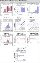
          The study analysed 43 eyes of 33 patients after three months and 35 eyes of 27 patients after 12 months. After 12 months, 100% of all eyes treated had ≤1.0 D and 97% ≤0.5 D of subjective residual astigmatism. Mean residual astigmatism was 0.27 D. 90% of all eyes were within one line of difference between UDVA and CDVA. SEQ Mean Absolute Error was 0.26 D and SEQ. Mean error was −0.08 ± 0.32 D. CI was 0.98 ± 0.2 D, and Index of Success, 0.20 ± 0.18 D.

          Conclusion

          The Castrop nomogram showed results that are comparable to or better than results presented in the literature for existing nomograms. Our results for astigmatic reduction are comparable to published results for TIOL implantation. It seems to be a predictable and safe measure to reduce manifest astigmatism.

          Related collections

          Most cited references43

          • Record: found
          • Abstract: found
          • Article: not found

          Contribution of posterior corneal astigmatism to total corneal astigmatism.

          To determine the contribution of posterior corneal astigmatism to total corneal astigmatism and the error in estimating total corneal astigmatism from anterior corneal measurements only using a dual-Scheimpflug analyzer. Cullen Eye Institute, Baylor College of Medicine, Houston, Texas, USA. Case series. Total corneal astigmatism was calculated using ray tracing, corneal astigmatism from simulated keratometry, anterior corneal astigmatism, and posterior corneal astigmatism, and the changes with age were analyzed. Vector analysis was used to assess the error produced by estimating total corneal astigmatism from anterior corneal measurements only. The study analyzed 715 corneas of 435 consecutive patients. The mean magnitude of posterior corneal astigmatism was -0.30 diopter (D). The steep corneal meridian was aligned vertically (60 to 120 degrees) in 51.9% of eyes for the anterior surface and in 86.6% for the posterior surface. With increasing age, the steep anterior corneal meridian tended to change from vertical to horizontal, while the steep posterior corneal meridian did not change. The magnitudes of anterior and posterior corneal astigmatism were correlated when the steeper anterior meridian was aligned vertically but not when it was aligned horizontally. Anterior corneal measurements underestimated total corneal astigmatism by 0.22 @ 180 and exceeded 0.50 D in 5% of eyes. Ignoring posterior corneal astigmatism may yield incorrect estimation of total corneal astigmatism. Selecting toric intraocular lenses based on anterior corneal measurements could lead to overcorrection in eyes that have with-the-rule astigmatism and undercorrection in eyes that have against-the-rule astigmatism. The authors received research support from Ziemer Group. In addition, Dr. Koch has a financial interest with Alcon Laboratories, Inc., Abbott Medical Optics, Inc., Calhoun Vision, Inc., NuLens, and Optimedica Corp. Copyright © 2012 ASCRS and ESCRS. Published by Elsevier Inc. All rights reserved.
            Bookmark
            • Record: found
            • Abstract: found
            • Article: not found

            Correcting astigmatism with toric intraocular lenses: effect of posterior corneal astigmatism.

            To evaluate the impact of posterior corneal astigmatism on outcomes with toric intraocular lenses (IOLs). Cullen Eye Institute, Baylor College of Medicine, Houston, Texas, USA. Case series. Corneal astigmatism was measured using 5 devices before and 3 weeks after cataract surgery. Toric IOL alignment was recorded at surgery and at the slitlamp 3 weeks postoperatively. The actual corneal astigmatism was calculated based on refractive astigmatism 3 weeks postoperatively and the effective toric power calculated with the Holladay 2 formula. The prediction error was calculated as the difference between the astigmatism measured by each device and the actual corneal astigmatism. Vector analysis was used in all calculations. With the IOLMaster, Lenstar, Atlas, manual keratometer, and Galilei (combined Placido-dual Scheimpflug analyzer), the mean prediction errors (D) were, respectively, 0.59 @ 89.7, 0.48 @ 91.2, 0.51 @ 78.7, 0.62 @ 97.2, and 0.57 @ 93.9 for with-the-rule (WTR) astigmatism (60 to 120 degrees), and 0.17 @ 86.2, 0.23 @ 77.7, 0.23 @ 91.4, 0.41 @ 58.4, and 0.12 @ 7.3 for against-the-rule (ATR) astigmatism (0 to 30 degrees and 150 to 180 degrees). In the WTR eyes, there were significant WTR prediction errors (0.5 to 0.6 diopters [D]) by all devices. In ATR eyes, WTR prediction errors were 0.2 to 0.3 D by all devices except the Placido-dual Scheimpflug analyzer (all P<.05 with Bonferroni correction). Corneal astigmatism was overestimated in WTR by all devices and underestimated in ATR by all except the Placido-dual Scheimpflug analyzer. A new toric IOL nomogram is proposed. Copyright © 2013 ASCRS and ESCRS. Published by Elsevier Inc. All rights reserved.
              Bookmark
              • Record: found
              • Abstract: found
              • Article: not found

              Analysis of biometry and prevalence data for corneal astigmatism in 23,239 eyes.

              To present and analyze biometry data sets and prevalence data for corneal astigmatism in a large population. High-volume eye surgery center, Castrop-Rauxel, Germany. Axial length (AL), corneal radii, anterior chamber depth (ACD), and horizontal corneal diameter (white-to-white [WTW] distance) were optically measured by partial coherence interferometry (IOLMaster). Patient data sets acquired between 2000 and 2006 were reviewed and analyzed. The study evaluated 23,239 data sets of 15,448 patients with a median age of 74 years. The mean values were as follows: AL, 23.43 mm +/- 1.51 (SD); corneal radius, 7.69 +/- 0.28 mm; WTW distance, 11.82 +/- 0.40 mm; and ACD, 3.11 +/- 0.43 mm. The ACD and axis of astigmatism were correlated with age. The AL, corneal radius, ACD, and WTW were correlated with one other. Eight percent of eyes had corneal astigmatism greater than 2.00 diopters (D), and 2.6% had more than 3.00 D. Astigmatism was with the rule (WTW) in 46.8% of eyes, against the rule in 34.4%, and oblique in 18.9%. High astigmatism was predominantly WTW. The results in this analysis might provide normative data for cataract patients and a useful reference for multiple purposes. The correlation of AL with corneal radius, ACD, and corneal diameter in normal eyes was not present in eyes with extreme myopia or hyperopia. Copyright (c) 2010 ASCRS and ESCRS. Published by Elsevier Inc. All rights reserved.
                Bookmark

                Author and article information

                Contributors
                jascha.wendelstein@kepleruniklinikum.at
                Journal
                Acta Ophthalmol
                Acta Ophthalmol
                10.1111/(ISSN)1755-3768
                AOS
                Acta Ophthalmologica
                John Wiley and Sons Inc. (Hoboken )
                1755-375X
                1755-3768
                24 February 2021
                December 2021
                : 99
                : 8 ( doiID: 10.1111/aos.v99.8 )
                : e1297-e1306
                Affiliations
                [ 1 ] Department of Ophthalmology and Optometry Kepler University Hospital GmbH Johannes Kepler University Linz Linz Austria
                [ 2 ] Augen‐und Laserklinik Castrop‐Rauxel Germany
                [ 3 ] VIROS – Vienna Institute for Research in Ocular Surgery Karl Landsteiner Institute Hanusch Hospital Vienna Austria
                Author notes
                [*] [* ] Correspondence:

                Jascha A. Wendelstein, MD

                Department of Ophthalmology and Optometry

                Kepler University Hospital GmbH

                Johannes Kepler University Linz

                Krankenhausstrasse 9

                4020 Linz

                Austria

                Tel: +43 (0)5 7680 83 ‐ 1048

                Fax: +43 (0)5 7680 83 ‐ 1822

                Email: jascha.wendelstein@ 123456kepleruniklinikum.at

                Author information
                https://orcid.org/0000-0003-4145-2559
                Article
                AOS14837
                10.1111/aos.14837
                9540120
                33629542
                16a88765-4e00-4b64-8b2f-87921bb9aa01
                © 2021 The Authors. Acta Ophthalmologica published by John Wiley & Sons Ltd on behalf of Acta Ophthalmologica Scandinavica Foundation.

                This is an open access article under the terms of the http://creativecommons.org/licenses/by-nc-nd/4.0/ License, which permits use and distribution in any medium, provided the original work is properly cited, the use is non‐commercial and no modifications or adaptations are made.

                History
                : 01 November 2020
                : 12 February 2021
                Page count
                Figures: 3, Tables: 3, Pages: 10, Words: 7807
                Funding
                Funded by: Bausch and Lomb , doi 10.13039/100007489;
                Categories
                Original Article
                Original Articles
                Custom metadata
                2.0
                December 2021
                Converter:WILEY_ML3GV2_TO_JATSPMC version:6.2.0 mode:remove_FC converted:07.10.2022

                Ophthalmology & Optometry
                arcuate incisions,astigmatism,castrop nomogram,corneal incisions,femtosecond laser,keratotomy

                Comments

                Comment on this article