70
views
0
recommends
+1 Recommend
0 collections
    4
    shares
      • Record: found
      • Abstract: found
      • Article: found
      Is Open Access

      Rasterstereographic measurement of scoliotic deformity

      research-article

      Read this article at

      Bookmark
          There is no author summary for this article yet. Authors can add summaries to their articles on ScienceOpen to make them more accessible to a non-specialist audience.

          Abstract

          Background

          Back surface topography has gained acceptance in recent decades. At the same time, the motivation to use this technique has increased. From the view of the patient, the cosmetic aspect has played and still plays a major role as it provides a comprehensive documentation of cosmetic impairment. From the view of the medical practitioner, the aspect of reducing X-ray exposures in diagnosis and follow-up has been dominant and still prevails. Meanwhile, new aspects have emerged: due to the consequent three-dimensional view of the scoliotic condition, treatment success can be visualized convincingly. Clinical diagnosis is supported by information otherwise not supplied by X-rays, such as when functional examinations and diagnostic tests are recorded.

          Methods

          Like rasterstereography, most techniques of actual back surface measurement refer to photogrammetry and the triangulation method. However, with respect to the particular clinical application, a wide spectrum of implementations exists. Applications in a clinic require high accuracy of measurement in a short time and comprehensive analysis providing data to be used to supplement and compare with radiographic data. This is exemplified by rasterstereography; the procedures of surface analysis and localization of landmarks using curvatures and the reconstruction of the spinal midline will be described.

          Orthopaedic relevance

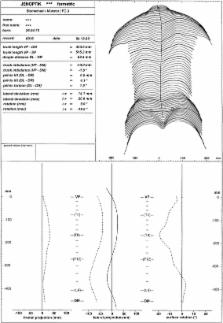
          Based on rasterstereographic analysis, different geometrical measures that characterize the back surface are given and underlying skeletal structures described. Furthermore, in analogy to radiological projection, a 3-D reconstruction of the spinal midline is visualized by a frontal and lateral projection, allowing comparison with pertinent X-rays.

          Conclusions

          Surface topography and, in particular, rasterstereography provide reliable and consistent results that may be used to reduce X-ray exposure. Unfortunately, the correlation of shape parameters with the radiological Cobb angle is poor. However, the wealth of additional applications substantially enhances the spectrum of clinical value.

          Related collections

          Most cited references57

          • Record: found
          • Abstract: not found
          • Article: not found

          Structured-light 3D surface imaging: a tutorial

          Jason Geng (2011)
            Bookmark
            • Record: found
            • Abstract: found
            • Article: not found

            An objective criterion for scoliosis screening.

            W Bunnell (1984)
            A specially designed inclinometer has been used to measure one part of the clinical deformity (asymmetry of the trunk) that is seen in scoliosis. This objective measurement provides one good guideline that can effectively determine, in surveys of children, whether or not further orthopaedic evaluation is needed. A minimum significant angle of trunk rotation of 5 degrees was shown by computer-analyzed data from 1,065 patients to be a good criterion for identifying curvatures of 20 degrees or more. The specificity of scoliosis screening in this manner has a projected false-negative rate of 0.1 per cent and a high degree of sensitivity. The method is simple, reliable, and inexpensive. It is easily taught to lay personnel who can be employed in scoliosis screening procedures.
              Bookmark
              • Record: found
              • Abstract: found
              • Article: not found

              Cancer mortality among women frequently exposed to radiographic examinations for spinal disorders.

              We studied cancer mortality in a cohort of 5,573 women with scoliosis and other spine disorders who were diagnosed between 1912 and 1965 and were exposed to frequent diagnostic X-ray procedures. Patients were identified from medical records in 14 orthopedic medical centers in the United States and followed for vital status and address through December 31, 2004, using publicly available regional, state and nationwide databases. Causes of death were obtained from death certificates or through linkage with the National Death Index (NDI). Statistical analyses included standardized mortality ratios (SMR = observed/expected) based on death rates for U.S. females and internal comparisons using Cox regression models with attained age as the time scale. Diagnostic radiation exposure was estimated from radiology files for over 137,000 procedures; estimated average cumulative radiation doses to the breast, lung, thyroid and bone marrow were 10.9, 4.1, 7.4 and 1.0 cGy, respectively. After a median follow-up period of 47 years, 1527 women died, including 355 from cancer. Cancer mortality was 8% higher than expected (95% CI = 0.97-1.20). Mortality from breast cancer was significantly elevated (SMR = 1.68; 95% CI: 1.38-2.02), whereas death rates from several other cancers were below expectation, in particular lung (SMR = 0.77), cervical (SMR = 0.31), and liver (SMR = 0.17). The excess relative risk (ERR) for breast cancer mortality increased significantly with 10-year lagged radiation dose to the breast (ERR/Gy = 3.9; 95% CI: 1.0-9.3).
                Bookmark

                Author and article information

                Contributors
                drerup@uni-muenster.de
                Journal
                Scoliosis
                Scoliosis
                Scoliosis
                BioMed Central (London )
                1748-7161
                12 December 2014
                12 December 2014
                2014
                : 9
                : 1
                : 22
                Affiliations
                Bundesfachschule für Orthopaedietechnik, Schliepstrasse 6-8, Dortmund, Germany
                Article
                22
                10.1186/s13013-014-0022-7
                4268794
                25520745
                1717eb3d-3171-4d26-8ec2-50876c06faa8
                © Drerup; licensee BioMed Central. 2014

                This is an Open Access article distributed under the terms of the Creative Commons Attribution License ( http://creativecommons.org/licenses/by/4.0), which permits unrestricted use, distribution, and reproduction in any medium, provided the original work is properly credited. The Creative Commons Public Domain Dedication waiver ( http://creativecommons.org/publicdomain/zero/1.0/) applies to the data made available in this article, unless otherwise stated.

                History
                : 21 October 2014
                : 26 November 2014
                Categories
                Review
                Custom metadata
                © The Author(s) 2014

                Orthopedics
                back surface,scoliosis,rasterstereography,photogrammetry,curvature map,symmetry line,anatomical landmarks,shape analysis,cobb angle

                Comments

                Comment on this article