57
views
0
recommends
+1 Recommend
0 collections
    0
    shares
      • Record: found
      • Abstract: found
      • Article: found
      Is Open Access

      Aberrant Brain Activity at Early Delay Stage Post-radiotherapy as a Biomarker for Predicting Neurocognitive Dysfunction Late-Delayed in Patients With Nasopharyngeal Carcinoma

      research-article

      Read this article at

      Bookmark
          There is no author summary for this article yet. Authors can add summaries to their articles on ScienceOpen to make them more accessible to a non-specialist audience.

          Abstract

          Background: Increasing evidence indicates that early radiation-induced subtle cerebral changes may be the precursors to permanent brain dysfunction at the late-delayed (LDS) post-radiotherapy (RT) stage. In this study, we aim to track the RT-related longitudinal brain activity in nasopharyngeal carcinoma (NPC) patients and to determine whether early abnormal brain activity can predict late neurocognitive dysfunction after RT.

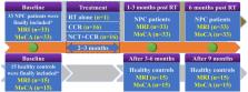
          Methods: Thirty-three NPC patients were finally included and longitudinally followed up at the following time points: prior to treatment initiation, early-delayed stage (EDS, 1–3 months), and LDS (six months) after RT. Fifteen comparable healthy controls (HCs) were finally included and followed up in parallel. Montreal Cognitive Assessment (MoCA) was used to assess the general cognitive function. Brain activity was recorded via resting-state fMRI and regional homogeneity (ReHo). A whole-brain voxel-wise-based one-way repeated-measure analysis of variance (ANOVA) was conducted to evaluate the longitudinal ReHo changes among the three time points for NPC patients and HCs, respectively. Results were reported at the significant level of a threshold of two-tailed voxel-wise P < 0.01 and cluster level P < 0.05 with Gaussian Random Field (GRF) correction. Finally, the efficacies of the aberrant ReHo at EDS for predicting the cognitive impairment at LDS in NPC patients were evaluated.

          Results: Significant differences were detected in ReHo among the three time points in NPC patients but not in HCs. Aberrant ReHo was distributed in the bilateral cerebellum, the right temporal lobe, and the left insular areas, which showed different dynamic changes patterns over time. Logistic regression model combining the mean ReHo, age, and irradiation dose on the bilateral temporal lobe had the highest diagnostic efficiency according to the area under the curve (AUC) score (AUC = 0.752, P = 0.023).

          Conclusions: The post-RT brain activity revealed by ReHo in NPC patients was dynamic, complex, and multifactorial. Furthermore, the combination of the aberrant ReHo at EDS, age, and irradiation dose may serve as a potential biomarker of the RT-induced cognitive impairments at LDS.

          Related collections

          Most cited references37

          • Record: found
          • Abstract: found
          • Article: not found

          Management of Nasopharyngeal Carcinoma: Current Practice and Future Perspective.

          Nasopharyngeal carcinoma of the undifferentiated subtype is endemic to southern China, and patient prognosis has improved significantly over the past three decades because of advances in disease management, diagnostic imaging, radiotherapy technology, and broader application of systemic therapy. Despite the excellent local control with modern radiotherapy, distant failure remains a key challenge. Advances in molecular technology have helped to decipher the molecular pathogenesis of nasopharyngeal carcinoma as well as its etiologic association with the Epstein-Barr virus. This in turn has led to the discovery of novel biomarkers and drug targets, rendering this cancer site a current focus for new drug development. This article reviews and appraises the key literature on the current management of nasopharyngeal carcinoma and future directions in clinical research.
            Bookmark
            • Record: found
            • Abstract: found
            • Article: not found

            Functional connectivity of the insula in the resting brain.

            The human insula is hidden in the depth of the cerebral hemisphere by the overlying frontal and temporal opercula, and consists of three cytoarchitectonically distinct regions: the anterior agranular area, posterior granular area, and the transitional dysgranular zone; each has distinct histochemical staining patterns and specific connectivity. Even though there are several studies reporting the functional connectivity of the insula with the cingulated cortex, its relationships with other brain areas remain elusive in humans. Therefore, we decided to use resting state functional connectivity to elucidate in details its connectivity, in terms of cortical and subcortical areas, and also of lateralization. We investigated correlations in BOLD fluctuations between specific regions of interest of the insula and other brain areas of right-handed healthy volunteers, on both sides of the brain. Our findings document two major complementary networks involving the ventral-anterior and dorsal-posterior insula: one network links the anterior insula to the middle and inferior temporal cortex and anterior cingulate cortex, and is primarily related to limbic regions which play a role in emotional aspects; the second links the middle-posterior insula to premotor, sensorimotor, supplementary motor and middle-posterior cingulate cortices, indicating a role for the insula in sensorimotor integration. The clear bipartition of the insula was confirmed by negative correlation analysis. Correlation maps are partially lateralized: the salience network, related to the ventral anterior insula, displays stronger connections with the anterior cingulate cortex on the right side, and with the frontal cortex on the left side; the posterior network has stronger connections with the superior temporal cortex and the occipital cortex on the right side. These results are in agreement with connectivity studies in primates, and support the use of resting state functional analysis to investigate connectivity in the living human brain. Copyright © 2010 Elsevier Inc. All rights reserved.
              Bookmark
              • Record: found
              • Abstract: found
              • Article: not found

              Test-retest reliabilities of resting-state FMRI measurements in human brain functional connectomics: a systems neuroscience perspective.

              Resting-state functional magnetic resonance imaging (RFMRI) enables researchers to monitor fluctuations in the spontaneous brain activities of thousands of regions in the human brain simultaneously, representing a popular tool for macro-scale functional connectomics to characterize normal brain function, mind-brain associations, and the various disorders. However, the test-retest reliability of RFMRI remains largely unknown. We review previously published papers on the test-retest reliability of voxel-wise metrics and conduct a meta-summary reliability analysis of seven common brain networks. This analysis revealed that the heteromodal associative (default, control, and attention) networks were mostly reliable across the seven networks. Regarding examined metrics, independent component analysis with dual regression, local functional homogeneity and functional homotopic connectivity were the three mostly reliable RFMRI metrics. These observations can guide the use of reliable metrics and further improvement of test-retest reliability for other metics in functional connectomics. We discuss the main issues with low reliability related to sub-optimal design and the choice of data processing options. Future research should use large-sample test-retest data to rectify both the within-subject and between-subject variability of RFMRI measurements and accelerate the application of functional connectomics.
                Bookmark

                Author and article information

                Contributors
                Journal
                Front Neurol
                Front Neurol
                Front. Neurol.
                Frontiers in Neurology
                Frontiers Media S.A.
                1664-2295
                16 July 2019
                2019
                : 10
                : 752
                Affiliations
                [1] 1Department of Medical Imaging, Sun Yat-sen University Cancer Center, State Key Laboratory of Oncology in South China, Collaborative Innovation Center for Cancer Medicine, Guangdong Key Laboratory of Nasopharyngeal Carcinoma Diagnosis and Therapy , Guangzhou, China
                [2] 2Department of Radiology, The Third Affiliated Hospital of Guangzhou Medical University, Guangzhou Medical University , Guangzhou, China
                [3] 3Department of Radiation Oncology, Sun Yat-sen University Cancer Center, State Key Laboratory of Oncology in South China, Collaborative Innovation Center for Cancer Medicine, Guangdong Key Laboratory of Nasopharyngeal Carcinoma Diagnosis and Therapy , Guangzhou, China
                [4] 4Shenzhen Kangning Hospital, Shenzhen Mental Health Center , Shenzhen, China
                Author notes

                Edited by: Feng Feng, Peking Union Medical College Hospital (CAMS), China

                Reviewed by: Bo Gao, Affiliated Hospital of Guizhou Medical University, China; Jordi A. Matias-Guiu, Servicio de Neurología, Hospital Clínico San Carlos, Spain

                *Correspondence: Xiaofei Lv lvxf@ 123456sysucc.org.cn

                This article was submitted to Applied Neuroimaging, a section of the journal Frontiers in Neurology

                †These authors have contributed equally to this work

                Article
                10.3389/fneur.2019.00752
                6660255
                192d43b3-a8d7-48db-93d9-214007e9025d
                Copyright © 2019 Yang, Lin, Li, Han, Li, Liu, Hou, Xie, Lv and Qiu.

                This is an open-access article distributed under the terms of the Creative Commons Attribution License (CC BY). The use, distribution or reproduction in other forums is permitted, provided the original author(s) and the copyright owner(s) are credited and that the original publication in this journal is cited, in accordance with accepted academic practice. No use, distribution or reproduction is permitted which does not comply with these terms.

                History
                : 30 March 2019
                : 27 June 2019
                Page count
                Figures: 4, Tables: 3, Equations: 0, References: 44, Pages: 10, Words: 7529
                Categories
                Neurology
                Original Research

                Neurology
                radiotherapy,radiation-induced injury,regional homogeneity,resting-state fmri,nasopharyngeal carcinoma,neurocognitive dysfunction

                Comments

                Comment on this article