8
views
0
recommends
+1 Recommend
0 collections
    0
    shares
      • Record: found
      • Abstract: found
      • Article: found
      Is Open Access

      An investigation into the effect of changing the computed tomography slice reconstruction interval on the spatial replication accuracy of three‐dimensional printed anatomical models constructed by fused deposition modelling

      research-article
      , BMedImg (Honours) 1 , 2 , , , MAppSc, MEd, FIR, SFHEA 2
      Journal of Medical Radiation Sciences
      John Wiley and Sons Inc.
      3D‐printing, computed tomography, fused deposition modelling, biofabrication, slice reconstruction interval

      Read this article at

      Bookmark
          There is no author summary for this article yet. Authors can add summaries to their articles on ScienceOpen to make them more accessible to a non-specialist audience.

          Abstract

          Introduction

          Three‐dimensional (3D) printed models can be constructed utilising computed tomography (CT) data. This project aimed to determine the effect of changing the slice reconstruction interval (SRI) on the spatial replication accuracy of 3D‐printed anatomical models constructed by fused deposition modelling (FDM).

          Methods

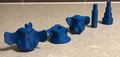
          Three bovine vertebrae and an imaging phantom were imaged using a CT scanner. The Queensland State Government’s Animal Care and Protection Act 2001 did not apply as no animals were harmed to carry out scientific activity. The data were reconstructed into SRIs of 0.1, 0.3, 0.5 and 1 mm and processed by software before 3D printing. Specimens and printed models were measured with calipers to calculate mean absolute error prior to statistical analysis.

          Results

          Mean absolute error from the original models for the 0.1, 0.3, 0.5 and 1 mm 3D‐printed models was 0.592 ± 0.396 mm, 0.598 ± 0.479 mm, 0.712 ± 0.498 mm and 0.933 ± 0.457 mm, respectively. Paired t‐tests ( P < 0.05) indicated a statistically significant difference between all original specimens and corresponding 3D‐printed models except the 0.1 mm vertebrae 2 ( P = 0.061), 0.3 mm phantom 1 ( P = 0.209) and 0.3 mm vertebrae 2 ( P = 0.097).

          Conclusion

          This study demonstrated that changing the SRI influences the spatial replication accuracy of 3D‐printed models constructed by FDM. Matching the SRI to the primary spatial resolution limiting factor of acquisition slice width or printer capabilities optimises replication accuracy.

          Abstract

          There are many applications of three‐dimensional printed models generated from computed tomography scan data. This project aimed to determine the effect of changing the slice reconstruction interval on the spatial replication accuracy of three‐dimensional printed anatomical models constructed by fused deposition modelling.

          Related collections

          Most cited references20

          • Record: found
          • Abstract: found
          • Article: not found

          The production of anatomical teaching resources using three-dimensional (3D) printing technology.

          The teaching of anatomy has consistently been the subject of societal controversy, especially in the context of employing cadaveric materials in professional medical and allied health professional training. The reduction in dissection-based teaching in medical and allied health professional training programs has been in part due to the financial considerations involved in maintaining bequest programs, accessing human cadavers and concerns with health and safety considerations for students and staff exposed to formalin-containing embalming fluids. This report details how additive manufacturing or three-dimensional (3D) printing allows the creation of reproductions of prosected human cadaver and other anatomical specimens that obviates many of the above issues. These 3D prints are high resolution, accurate color reproductions of prosections based on data acquired by surface scanning or CT imaging. The application of 3D printing to produce models of negative spaces, contrast CT radiographic data using segmentation software is illustrated. The accuracy of printed specimens is compared with original specimens. This alternative approach to producing anatomically accurate reproductions offers many advantages over plastination as it allows rapid production of multiple copies of any dissected specimen, at any size scale and should be suitable for any teaching facility in any country, thereby avoiding some of the cultural and ethical issues associated with cadaver specimens either in an embalmed or plastinated form.
            Bookmark
            • Record: found
            • Abstract: not found
            • Article: not found

            Measuring and Establishing the Accuracy and Reproducibility of 3D Printed Medical Models

              Bookmark
              • Record: found
              • Abstract: found
              • Article: not found

              Accuracy of medical models made by additive manufacturing (rapid manufacturing).

              Additive manufacturing (AM) is being increasingly used for producing medical models. The accuracy of these models varies between different materials, AM technologies and machine runs.
                Bookmark

                Author and article information

                Contributors
                searle.j.benjamin@gmail.com
                Journal
                J Med Radiat Sci
                J Med Radiat Sci
                10.1002/(ISSN)2051-3909
                JMRS
                Journal of Medical Radiation Sciences
                John Wiley and Sons Inc. (Hoboken )
                2051-3895
                2051-3909
                13 February 2020
                March 2020
                : 67
                : 1 , Optimisation in radiation therapy and medical imaging service provision and education ( doiID: 10.1002/jmrs.v67.1 )
                : 43-53
                Affiliations
                [ 1 ] Redcliffe Hospital Redcliffe QLD Australia
                [ 2 ] School of Clinical Sciences Faculty of Health Queensland University of Technology Brisbane QLD Australia
                Author notes
                [*] [* ] Correspondence

                Benjamin Searle, School of Clinical Sciences, Faculty of Health, Queensland University of Technology, Queensland, Australia. Tel: +61401175483; Fax: +6173138152; Email: searle.j.benjamin@ 123456gmail.com

                Author information
                https://orcid.org/0000-0003-1227-0721
                https://orcid.org/0000-0002-8353-4977
                Article
                JMRS382
                10.1002/jmrs.382
                7063250
                32056391
                1a13958c-6a77-40ff-bf79-84ee911da88f
                © 2020 The Authors. Journal of Medical Radiation Sciences published by John Wiley & Sons Australia, Ltd on behalf of Australian Society of Medical Imaging and Radiation Therapy and New Zealand Institute of Medical Radiation Technology

                This is an open access article under the terms of the http://creativecommons.org/licenses/by-nc/4.0/ License, which permits use, distribution and reproduction in any medium, provided the original work is properly cited and is not used for commercial purposes.

                History
                : 06 September 2019
                : 07 January 2020
                : 07 January 2020
                Page count
                Figures: 6, Tables: 5, Pages: 11, Words: 7700
                Categories
                Original Article
                Original Articles
                Custom metadata
                2.0
                March 2020
                Converter:WILEY_ML3GV2_TO_JATSPMC version:5.7.7 mode:remove_FC converted:10.03.2020

                3d‐printing,computed tomography,fused deposition modelling,biofabrication,slice reconstruction interval

                Comments

                Comment on this article