7
views
0
recommends
+1 Recommend
0 collections
    0
    shares
      • Record: found
      • Abstract: found
      • Article: found
      Is Open Access

      Bioactivity guided isolation of cytotoxic terpenoids and steroids from Premna serratifolia

      research-article

      Read this article at

      Bookmark
          There is no author summary for this article yet. Authors can add summaries to their articles on ScienceOpen to make them more accessible to a non-specialist audience.

          Abstract

          Context: Despite several phytochemical studies of Premna serratifolia Linn. (Verbenaceae), the isolation of active constituents of this plant remains to be explored.

          Objective: The study isolates cytotoxic terpenoids and steroids from the leaves of Premna serratifolia.

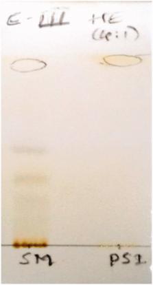
          Materials and methods: Unsaponifiable matter of hexane soluble fraction obtained from methanol extract was subjected to isolation by column chromatography and preparative TLC. Three compounds PS-01 A, PS-01B and PS-02 A were isolated. PS-01 A and PS-01B were identified by comparative TLC with authentic marker compounds followed by NMR analysis. Further PS-01B was analyzed by HR-GCMS. PS-02 A was subjected to HR-LCMS. All isolated compounds/fractions were evaluated for cytotoxic activity by BSL bioassay and using cell lines A549, HepG2 and L6.

          Results: Three compounds were isolated from the leaf extract by bioactivity-guided fractionation. Two of which, namely, PS-01 A (oleanolic acid) and PS-02 A (unknown) were found to be terpenoids and PS-01B was identified as steroid (stigmasterol). PS-02 A compound is to be purified and characterized further. All three compounds PS-01 A, PS-01B, PS-02 A showed cytotoxicity by BSL bioassay (LC 50 value of 54.49, 30.83, 16.32 ppm, respectively) and by cell line study where isolate PS-02 A has shown more cytotoxicity with LC 50 values of 66.77 and 53.72 μg/mL with A549 and HepG2 cells, respectively, when compared with other isolates.

          Conclusion: Bioactivity guided fractionation of Premna serratifolia leaves succeeded into isolation of two terpenoids and one steroid compound with significant cytotoxic activity. Here we report the isolation of these cytotoxic terpenoids/steroids from this plant for the first time which could be developed as anticancer agents.

          Related collections

          Most cited references13

          • Record: found
          • Abstract: found
          • Article: not found

          Terpenoids as potential chemopreventive and therapeutic agents in liver cancer.

          Despite significant advances in medicine, liver cancer, predominantly hepatocellular carcinoma remains a major cause of death in the United States as well as the rest of the world. As limited treatment options are currently available to patients with liver cancer, novel preventive control and effective therapeutic approaches are considered to be reasonable and decisive measures to combat this disease. Several naturally occurring dietary and non-dietary phytochemicals have shown enormous potential in the prevention and treatment of several cancers, especially those of the gastrointestinal tract. Terpenoids, the largest group of phytochemicals, traditionally used for medicinal purposes in India and China, are currently being explored as anticancer agents in clinical trials. Terpenoids (also called "isoprenoids") are secondary metabolites occurring in most organisms, particularly plants. More than 40 000 individual terpenoids are known to exist in nature with new compounds being discovered every year. A large number of terpenoids exhibit cytotoxicity against a variety of tumor cells and cancer preventive as well as anticancer efficacy in preclinical animal models. This review critically examines the potential role of naturally occurring terpenoids, from diverse origins, in the chemoprevention and treatment of liver tumors. Both in vitro and in vivo effects of these agents and related cellular and molecular mechanisms are highlighted. Potential challenges and future directions involved in the advancement of these promising natural compounds in the chemoprevention and therapy of human liver cancer are also discussed.
            Bookmark
            • Record: found
            • Abstract: found
            • Article: found
            Is Open Access

            Isolation and evaluation of anticancer efficacy of stigmasterol in a mouse model of DMBA-induced skin carcinoma

            Stigmasterol (99.9% pure) was isolated from Azadirachta indica and its chemopreventive effect on 7,12-dimethylbenz[a]anthracene (DMBA)-induced skin cancer was investigated in Swiss albino mice. Skin tumors were induced by topical application of DMBA and promoted by croton oil. To assess the chemopreventive potential of stigmasterol, it was orally administered at a concentration of 200 mg/kg and 400 mg/kg three times weekly for 16 weeks. Reduction in tumor size and cumulative number of papillomas were seen as a result of treatment with stigmasterol. The average latency period was significantly increased as compared with the carcinogen-treated control. Stigmasterol induced a significant decrease in the activity of serum enzymes, such as aspartate aminotransferase, alanine aminotransferase, alkaline phosphatase, and bilirubin as compared with the control. Stigmasterol significantly increased glutathione, superoxide dismutase, and catalase as compared with the control. Elevated levels of lipid peroxide and DNA damage in the control group were significantly inhibited by administration of stigmasterol. From the present study, it can be inferred that stigmasterol has chemopreventive activity in an experimental model of cancer. This chemopreventive activity may be linked to the oxidative stress of stigmasterol. The antigenotoxic properties of stigmasterol are also likely to contribute to its chemopreventive action.
              Bookmark
              • Record: found
              • Abstract: found
              • Article: found
              Is Open Access

              Anticancer and apoptotic activities of oleanolic acid are mediated through cell cycle arrest and disruption of mitochondrial membrane potential in HepG2 human hepatocellular carcinoma cells

              Hepatocellular carcinoma (HCC) is an aggressive form of cancer, with high rates of morbidity and mortality, a poor prognosis and limited therapeutic options. The objective of the present study was to demonstrate the anticancer activity of oleanolic acid in HepG2 human HCC cells. Cell viability was evaluated using an MTT assay, following administration of various doses of oleanolic acid. The effect of oleanolic acid on cell cycle phase distribution and mitochondrial membrane potential was evaluated using flow cytometry with propidium iodide and rhodamine-123 DNA-binding cationic fluorescent dyes. Fluorescence microscopy was employed to detect morphological changes in HepG2 cells following oleanolic acid treatment. The results revealed that oleanolic acid induced a dose-dependent, as well as time-dependent inhibition in the growth of HepG2 cancer cells. Following acridine orange and ethidium bromide staining, treatment with various doses (0, 5, 25 and 50 µM) of oleanolic acid induced typical morphological changes associated with apoptosis, including cell shrinkage, membrane blebbing, nuclear condensation and apoptotic body formation. Cell cycle analysis revealed that oleanolic acid induced cell cycle arrest in HepG2 cells at the sub-G1 (apoptotic) phase of the cell cycle, in a dose-dependent manner. Staining with Annexin V-fluorescein isothiocyanate and propidium iodide revealed that apoptosis occurred early in these cells. Oleanolic acid treatment also resulted in fragmentation of nuclear DNA in a dose-dependent manner, producing the typical features of DNA laddering on an agarose gel. The results also demonstrated that oleanolic acid treatment resulted in a potent loss of mitochondrial membrane potential, which also occurred in a dose-dependent manner. Therefore, oleanolic acid may be used as a therapeutic agent in the treatment of human HCC.
                Bookmark

                Author and article information

                Journal
                Pharm Biol
                Pharm Biol
                IPHB
                iphb20
                Pharmaceutical Biology
                Taylor & Francis
                1388-0209
                1744-5116
                2017
                19 March 2017
                : 55
                : 1
                : 1375-1379
                Affiliations
                Department of Pharmaconosy, KLE University’s College of Pharmacy , Belagavi, India
                Author notes
                CONTACT Kirankumar Hullatti drhullatti@ 123456klepharm.edu Department of Pharmaconosy, KLE University’s College of Pharmacy , Neharu Nagar, Belagavi, 590 010, India
                Article
                1301491
                10.1080/13880209.2017.1301491
                6130453
                28317412
                1a9cd721-189f-4670-8a3e-1668ff4bf98d
                © 2017 The Author(s). Published by Informa UK Limited, trading as Taylor & Francis Group.

                This is an Open Access article distributed under the terms of the Creative Commons Attribution License ( http://creativecommons.org/licenses/by/4.0/), which permits unrestricted use, distribution, and reproduction in any medium, provided the original work is properly cited.

                History
                : 04 July 2016
                : 04 February 2017
                : 28 February 2017
                Page count
                Pages: 5
                Categories
                Research Article

                fractionation,column chromatography,preparative tlc,bsl bioassay,cell line studies

                Comments

                Comment on this article

                scite_

                Similar content135

                Cited by4

                Most referenced authors104