168
views
0
recommends
+1 Recommend
0 collections
    0
    shares
      • Record: found
      • Abstract: found
      • Article: found
      Is Open Access

      Gliomas and the vascular fragility of the blood brain barrier

      review-article

      Read this article at

      Bookmark
          There is no author summary for this article yet. Authors can add summaries to their articles on ScienceOpen to make them more accessible to a non-specialist audience.

          Abstract

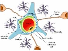
          Astrocytes, members of the glial family, interact through the exchange of soluble factors or by directly contacting neurons and other brain cells, such as microglia and endothelial cells. Astrocytic projections interact with vessels and act as additional elements of the Blood Brain Barrier (BBB). By mechanisms not fully understood, astrocytes can undergo oncogenic transformation and give rise to gliomas. The tumors take advantage of the BBB to ensure survival and continuous growth. A glioma can develop into a very aggressive tumor, the glioblastoma (GBM), characterized by a highly heterogeneous cell population (including tumor stem cells), extensive proliferation and migration. Nevertheless, gliomas can also give rise to slow growing tumors and in both cases, the afflux of blood, via BBB is crucial. Glioma cells migrate to different regions of the brain guided by the extension of blood vessels, colonizing the healthy adjacent tissue. In the clinical context, GBM can lead to tumor-derived seizures, which represent a challenge to patients and clinicians, since drugs used for its treatment must be able to cross the BBB. Uncontrolled and fast growth also leads to the disruption of the chimeric and fragile vessels in the tumor mass resulting in peritumoral edema. Although hormonal therapy is currently used to control the edema, it is not always efficient. In this review we comment the points cited above, considering the importance of the BBB and the concerns that arise when this barrier is affected.

          Related collections

          Most cited references107

          • Record: found
          • Abstract: not found
          • Article: not found

          CBTRUS statistical report: primary brain and central nervous system tumors diagnosed in the United States in 2005-2009.

            Bookmark
            • Record: found
            • Abstract: found
            • Article: not found

            Circulating microRNA in body fluid: a new potential biomarker for cancer diagnosis and prognosis.

            In the past several years, the importance of microRNA (miRNA) in cancer cells has been recognized. Proper control of miRNA expression is essential for maintaining a steady state of the cellular machinery. Recently, it was discovered that extracellular miRNAs circulate in the blood of both healthy and diseased patients, although ribonuclease is present in both plasma and serum. Most of the circulating miRNAs are included in lipid or lipoprotein complexes, such as apoptotic bodies, microvesicles, or exosomes, and are, therefore, highly stable. The existence of circulating miRNAs in the blood of cancer patients has raised the possibility that miRNAs may serve as a novel diagnostic marker. However, the secretory mechanism and biological function, as well as the meaning of the existence of extracellular miRNAs, remain largely unclear. In this review, we summarize the usefulness of circulating miRNA for cancer diagnosis, prognosis, and therapeutics. Furthermore, we propose a mechanism for the secretion and incorporation of miRNA into the cells. © 2010 Japanese Cancer Association.
              Bookmark
              • Record: found
              • Abstract: found
              • Article: not found

              Glial regulation of the cerebral microvasculature.

              The brain is a heterogeneous organ with regionally varied and constantly changing energetic needs. Blood vessels in the brain are equipped with control mechanisms that match oxygen and glucose delivery through blood flow with the local metabolic demands that are imposed by neural activity. However, the cellular bases of this mechanism have remained elusive. A major advance has been the demonstration that astrocytes, cells with extensive contacts with both synapses and cerebral blood vessels, participate in the increases in flow evoked by synaptic activity. Their organization in nonoverlapping spatial domains indicates that they are uniquely positioned to shape the spatial distribution of the vascular responses that are evoked by neural activity. Astrocytic calcium is an important determinant of microvascular function and may regulate flow independently of synaptic activity. The involvement of astrocytes in neurovascular coupling has broad implications for the interpretation of functional imaging signals and for the understanding of brain diseases that are associated with neurovascular dysfunction.
                Bookmark

                Author and article information

                Contributors
                Journal
                Front Cell Neurosci
                Front Cell Neurosci
                Front. Cell. Neurosci.
                Frontiers in Cellular Neuroscience
                Frontiers Media S.A.
                1662-5102
                12 December 2014
                2014
                : 8
                : 418
                Affiliations
                [1] 1Instituto Estadual do Cérebro Paulo Niemeyer, Rua do Rezende Rio de Janeiro, Brazil
                [2] 2Laboratório de Morfogênese Celular, Instituto de Ciências Biomédicas da, Universidade Federal do Rio de Janeiro Rio de Janeiro, Brazil
                [3] 3Programa de Pós-Graduação em Odontologia, Escola de Ciências da Saúde (ECS), Universidade do Grande Rio (UNIGRANRIO) Duque de Caxias, Brazil
                [4] 4Centro de Neurociência e Biologia Celular, Faculdade de Medicina, Universidade de Coimbra Coimbra, Portugal
                [5] 5Instituto de Ciências Biológicas e da Saúde, Universidade Federal de Alagoas, Maceió Alagoas, Brazil
                Author notes

                Edited by: Ramon Santos El-Bachá, Universidade Federal da Bahia, Brazil

                Reviewed by: Ulkan Kilic, Bezmialem Vakif University, Turkey; Andrey Turchinovich, German Cancer Research Center, Germany

                *Correspondence: Vivaldo Moura-Neto, Instituto Estadual do Cérebro Paulo Niemeyer, Rua do Rezende, 156, Rio de Janeiro RJ 20231-092, Brazil e-mail: vivaldo@ 123456icb.ufrj.br ; vivaldomouraneto@ 123456globo.com

                These authors have contributed equally to this work.

                This article was submitted to the journal Frontiers in Cellular Neuroscience.

                Article
                10.3389/fncel.2014.00418
                4264502
                25565956
                1c005f45-40f3-494c-be7d-85010a2b9e7c
                Copyright © 2014 Dubois, Campanati, Righy, D’Andrea-Meira, Spohr, Porto-Carreiro, Pereira, Balça-Silva, Kahn, DosSantos, de Almeida Rabello Oliveira, Ximenes-da-Silva, Lopes, Faveret, Gasparetto and Moura-Neto.

                This is an open-access article distributed under the terms of the Creative Commons Attribution License (CC BY). The use, distribution and reproduction in other forums is permitted, provided the original author(s) or licensor are credited and that the original publication in this journal is cited, in accordance with accepted academic practice. No use, distribution or reproduction is permitted which does not comply with these terms.

                History
                : 21 July 2014
                : 18 November 2014
                Page count
                Figures: 3, Tables: 0, Equations: 0, References: 145, Pages: 13, Words: 11339
                Categories
                Neuroscience
                Review Article

                Neurosciences
                glioblastoma,blood-brain barrier,mirna,exosomes,neural stem cells,tumor-related epileptic seizures,brain tumor related edema

                Comments

                Comment on this article