25
views
0
recommends
+1 Recommend
0 collections
    0
    shares
      • Record: found
      • Abstract: found
      • Article: found
      Is Open Access

      Impact of Age-Dependent Adventitia Inflammation on Structural Alteration of Abdominal Aorta in Hyperlipidemic Mice

      research-article

      Read this article at

      Bookmark
          There is no author summary for this article yet. Authors can add summaries to their articles on ScienceOpen to make them more accessible to a non-specialist audience.

          Abstract

          Background

          The adventitia is suggested to contribute to vascular remodeling; however, the site-selective inflammatory responses in association with the development of atherosclerosis remain to be elucidated.

          Methods and Results

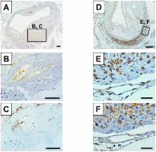
          Wild-type or apolipoprotein E knockout male C57BL/6J background mice were fed standard chow for 16, 32, and 52 weeks, and the morphology of the aortic arch, descending aorta, and abdominal aorta was compared. Atheromatous plaque formation progressed with age, particularly in the aortic arch and abdominal aorta but not in the descending aorta. In addition, we found that the numbers of macrophages, T-lymphocytes, and microvessels, assessed by anti-F4/80, CD3, and CD31 antibodies, were higher in the adventitia of the abdominal aorta at 52 weeks. These numbers were positively correlated with plaque formation, but negatively correlated with elastin content, resulting in the enlargement of the total vessel area. In aortic tissues, interleukin-6 levels increased in the atheromatous plaque with age, whereas the level of regulated on activation, normal T cell expressed and secreted (RANTES) increased with age, and compared with other sites, it was particularly distributed in inflammatory cells in the adventitia of the abdominal aorta.

          Conclusion

          This study suggests that adventitial inflammation contributes to the age-dependent structural alterations, and that the activation/inactivation of cytokines/chemokines is involved in the process.

          Related collections

          Most cited references27

          • Record: found
          • Abstract: not found
          • Article: not found

          Arterial and cardiac aging: major shareholders in cardiovascular disease enterprises: Part I: aging arteries: a "set up" for vascular disease.

            Bookmark
            • Record: found
            • Abstract: found
            • Article: not found

            Increased expression of matrix metalloproteinases and matrix degrading activity in vulnerable regions of human atherosclerotic plaques.

            Dysregulated extracellular matrix (ECM) metabolism may contribute to vascular remodeling during the development and complication of human atherosclerotic lesions. We investigated the expression of matrix metalloproteinases (MMPs), a family of enzymes that degrade ECM components in human atherosclerotic plaques (n = 30) and in uninvolved arterial specimens (n = 11). We studied members of all three MMP classes (interstitial collagenase, MMP-1; gelatinases, MMP-2 and MMP-9; and stromelysin, MMP-3) and their endogenous inhibitors (TIMPs 1 and 2) by immunocytochemistry, zymography, and immunoprecipitation. Normal arteries stained uniformly for 72-kD gelatinase and TIMPs. In contrast, plaques' shoulders and regions of foam cell accumulation displayed locally increased expression of 92-kD gelatinase, stromelysin, and interstitial collagenase. However, the mere presence of MMP does not establish their catalytic capacity, as the zymogens lack activity, and TIMPs may block activated MMPs. All plaque extracts contained activated forms of gelatinases determined zymographically and by degradation of 3H-collagen type IV. To test directly whether atheromata actually contain active matrix-degrading enzymes in situ, we devised a method which allows the detection and microscopic localization of MMP enzymatic activity directly in tissue sections. In situ zymography revealed gelatinolytic and caseinolytic activity in frozen sections of atherosclerotic but not of uninvolved arterial tissues. The MMP inhibitors, EDTA and 1,10-phenanthroline, as well as recombinant TIMP-1, reduced these activities which colocalized with regions of increased immunoreactive MMP expression, i.e., the shoulders, core, and microvasculature of the plaques. Focal overexpression of activated MMP may promote destabilization and complication of atherosclerotic plaques and provide novel targets for therapeutic intervention.
              Bookmark
              • Record: found
              • Abstract: found
              • Article: not found

              Endothelial cell senescence in human atherosclerosis: role of telomere in endothelial dysfunction.

              The functional changes associated with cellular senescence may be involved in human aging and age-related vascular disorders. We have shown the important role of telomere and telomerase in vascular cell senescence in vitro. Progressive telomere shortening in vivo has been observed in the regions susceptible to atherosclerosis, implying contributions to atherogenesis. However, whether senescent vascular cells are present in the vasculature and contribute to the pathogenesis of atherosclerosis remains unclear. Senescence-associated beta-galactosidase (beta-gal) activity was examined in the coronary arteries and the internal mammary arteries retrieved from autopsied individuals who had had ischemic heart diseases. Strong beta-gal stainings were observed in atherosclerotic lesions of the coronary arteries but not in the internal mammary arteries. An immunohistochemical analysis using anti-factor VIII antibody demonstrated that beta-gal stained cells are vascular endothelial cells. To determine whether endothelial cell senescence causes endothelial dysfunction, we induced senescence in human aortic endothelial cells (HAECs) by inhibiting telomere function and examined the expression of intercellular adhesion molecule (ICAM)-1 and endothelial nitric oxide synthase (eNOS) activity. Senescent HAECs exhibited increased ICAM-1 expression and decreased eNOS activity, both of which are alterations implicated in atherogenesis. In contrast, introduction of telomerase catalytic component significantly extended the life span and inhibited the functional alterations associated with senescence in HAECs. Vascular endothelial cells with senescence-associated phenotypes are present in human atherosclerotic lesions, and endothelial cell senescence induced by telomere shortening may contribute to atherogenesis.
                Bookmark

                Author and article information

                Contributors
                Role: Editor
                Journal
                PLoS One
                PLoS ONE
                plos
                plosone
                PLoS ONE
                Public Library of Science (San Francisco, USA )
                1932-6203
                2014
                25 August 2014
                : 9
                : 8
                : e105739
                Affiliations
                [1 ]Department of Internal Medicine, Circulatory and Body Fluid Regulation, Faculty of Medicine, University of Miyazaki, Miyazaki, Japan
                [2 ]Department of Pathology, Faculty of Medicine, University of Miyazaki, Miyazaki, Japan
                [3 ]Department of Internal Medicine, Koga General Hospital, Miyazaki, Japan
                University of Pittsburgh School of Medicine, United States of America
                Author notes

                Competing Interests: The authors have declared that no competing interests exist.

                Conceived and designed the experiments: SS TT. Performed the experiments: SS TT. Analyzed the data: SS TT. Contributed reagents/materials/analysis tools: SS TT KH TI YA KK. Wrote the paper: SS TT. Criticized and discussed experimental results: SS TT KH TI YA KK. Participated in proofreading and finalizing manuscript: SS TT KH TI YA KK.

                Article
                PONE-D-14-00962
                10.1371/journal.pone.0105739
                4143271
                25153991
                1c0c62de-c3d4-4dba-8faf-d8f6e16512a7
                Copyright @ 2014

                This is an open-access article distributed under the terms of the Creative Commons Attribution License, which permits unrestricted use, distribution, and reproduction in any medium, provided the original author and source are credited.

                History
                : 20 January 2014
                : 28 July 2014
                Page count
                Pages: 9
                Funding
                This study was supported by Grant-in-Aid for Scientific Research (C) (20590830) from the Ministry of Education, Culture, Sport, Science and Technology, Japan; Grant-in-Aid for the Japan Cardiovascular Research Foundation; Mitsubishi Pharma Research Foundation; Takeda Science Foundation; SENSHIN Medical Research Foundation. The funders had no role in study design, data collection and analysis, decision to publish, or preparation of the manuscript.
                Categories
                Research Article
                Medicine and Health Sciences
                Vascular Medicine
                Aortic Diseases
                Atherosclerosis
                Cardiology

                Uncategorized
                Uncategorized

                Comments

                Comment on this article