13
views
0
recommends
+1 Recommend
0 collections
    0
    shares
      • Record: found
      • Abstract: found
      • Article: found
      Is Open Access

      MR-arthrography and CT-arthrography in sports-related glenolabral injuries: a matched descriptive illustration

      review-article

      Read this article at
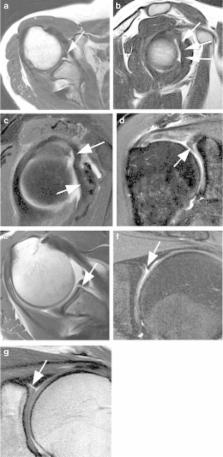
      Bookmark
          There is no author summary for this article yet. Authors can add summaries to their articles on ScienceOpen to make them more accessible to a non-specialist audience.

          Abstract

          The combination of a large range of motion and insufficient bony stabilization makes the glenohumeral joint susceptible to injuries including dislocation in young athletes. Magnetic resonance arthrography (MR-arthrography) and computed tomography arthrography (CT-arthrography) play an important role in the preoperative workup of labroligametous injuries. This paper illustrates MR-arthrography and CT-arthrography findings acquired at the same time on the same subjects to illustrate common causes and sequelae of shoulder instability.

          Teaching Points

          MR-arthrography and CT-arthrography are equivalent for SLAP and full-thickness rotator cuff tears .

          CT-arthrography is superior in evaluating osseous defects and cartilage surface lesions.

          MR-arthrography is superior in evaluating intrasubstance and extra-articular tendinous injuries .

          Related collections

          Most cited references25

          • Record: found
          • Abstract: found
          • Article: not found

          SLAP lesions of the shoulder.

          A specific pattern of injury to the superior labrum of the shoulder was identified arthroscopically in twenty-seven patients included in a retrospective review of more than 700 shoulder arthroscopies performed at our institution. The injury of the superior labrum begins posteriorly and extends anteriorly, stopping before or at the mid-glenoid notch and including the "anchor" of the biceps tendon to the labrum. We have labeled this injury a "SLAP lesion" (Superior Labrum Anterior and Posterior). There were 23 males and four females with an average age of 37.5 years. Time from injury to surgery averaged 29.3 months. The most common mechanism of injury was a compression force to the shoulder, usually as the result of a fall onto an outstretched arm, with the shoulder positioned in abduction and slight forward flexion at the time of the impact. The most common clinical complaints were pain, greater with overhead activity, and a painful "catching" or "popping" in the shoulder. No imaging test accurately defined the superior labral pathology preoperatively. We divided the superior labrum pathology into four distinct types. Treatment was performed arthroscopically based on the type of SLAP lesion noted at the time of surgery. The SLAP lesion, which has not been previously described, can be diagnosed only arthroscopically and may be treated successfully by arthroscopic techniques alone in many patients.
            Bookmark
            • Record: found
            • Abstract: found
            • Article: not found

            The incidence and characteristics of shoulder instability at the United States Military Academy.

            The literature provides little information detailing the incidence of traumatic shoulder instability in young, healthy athletes. Shoulder instability is common in young athletes. Descriptive epidemiologic study. We prospectively captured all traumatic shoulder instability events at the United States Military Academy between September 1, 2004, and May 31, 2005. Throughout this period, all new traumatic shoulder instability events were evaluated with physical examination, plain radiographs, and magnetic resonance imaging. Instability events were classified according to direction, chronicity, and type (subluxation or dislocation). Subject demographics, mechanism of injury, and sport were evaluated. Among 4141 students, 117 experienced new traumatic shoulder instability events during the study period; 11 experienced multiple events. The mean age of these 117 subjects was 20.0 years; 101 students were men (86.3%), and 16 were women (13.7%). The 1-year incidence proportion was 2.8%. The male incidence proportion was 2.9% and the female incidence proportion was 2.5%. Eighteen events were dislocations (15.4%), and 99 were subluxations (84.6%). Of the 99 subluxations, 45 (45.5%) were primary events, while 54 (54.5%) were recurrent. Of the 18 dislocations, 12 (66.7%) were primary events, while 6 (33.3%) were recurrent. The majority of the 117 events were anterior in nature (80.3%), while 12 (10.3%) were posterior, and 11 (9.4%) were multidirectional. Forty-four percent (43.6%) of the instability events experienced were as a result of contact injuries, while 41.0% were a result of noncontact injuries, including 9 subluxations caused by missed punches during boxing; information was unavailable for the remaining 15%. Glenohumeral instability is a common injury in this population, with subluxations comprising 85% of instability events.
              Bookmark
              • Record: found
              • Abstract: found
              • Article: not found

              Glenoid labrum tears related to the long head of the biceps.

              Tears of the glenoid labrum were observed in 73 baseball pitchers and other throwing athletes who underwent arthroscopic examination of the dominant shoulder. Most of the tears were located over the anterosuperior portion of the glenoid labrum near the origin of the tendon of the long head of the biceps muscle into the glenoid. At arthroscopy, the tendon of the long head of the biceps appeared to originate through and be continuous with the superior portion of the glenoid labrum. In many cases it appeared to have pulled the anterosuperior portion of the labrum off the glenoid. This observation was verified at arthroscopy by viewing the origin of the biceps tendon into the glenoid labrum as the muscle was electrically stimulated. With stimulation of the muscle, the tendinous portion became quite taut, particularly near its attachment to the glenoid labrum, and actually lifted the labrum off the glenoid. Three-dimensional high-speed cinematography with computer analysis revealed that the moment acting about the elbow joint to extend the joint through an arc of about 50 degrees was in excess of 600 inch-pounds. The extremely high velocity of elbow extension which is generated must be decelerated through the final 30 degrees of elbow extension. Of the muscles of the arm that provide the large deceleration forces in the follow-through phase of throwing, only the biceps brachii traverses both the elbow joint and the shoulder joint. Additional forces are generated in the biceps tendon in its function as a "shunt" muscle to stabilize the glenohumeral joint during the throwing act.(ABSTRACT TRUNCATED AT 250 WORDS)
                Bookmark

                Author and article information

                Contributors
                mohamedjarraya@gmail.com
                Journal
                Insights Imaging
                Insights Imaging
                Insights into Imaging
                Springer Berlin Heidelberg (Berlin/Heidelberg )
                1869-4101
                8 January 2016
                8 January 2016
                April 2016
                : 7
                : 2
                : 167-177
                Affiliations
                [ ]Aspetar Orthopaedic and Sports Medicine Hospital, Doha, Qatar
                [ ]Department of Radiology, Boston University School of Medicine, 820 Harrison Avenue, FGH Building, 3rd floor, Boston, MA USA
                [ ]Department of Radiology, Mercy Catholic Medical Center, Darby, PA USA
                [ ]Department of Radiology, University of Erlangen-Nuremberg, Erlangen, Germany
                [ ]Department of Radiology, Massachusetts General Hospital, Harvard Medical School, Boston, MA USA
                Author information
                http://orcid.org/0000-0002-8095-0174
                Article
                462
                10.1007/s13244-015-0462-5
                4805613
                26746976
                1c8c88ce-d950-4d37-8212-6793e9ef2cfa
                © The Author(s) 2016

                Open Access This article is distributed under the terms of the Creative Commons Attribution 4.0 International License (http://creativecommons.org/licenses/by/4.0/), which permits unrestricted use, distribution, and reproduction in any medium, provided you give appropriate credit to the original author(s) and the source, provide a link to the Creative Commons license, and indicate if changes were made.

                History
                : 28 September 2015
                : 14 December 2015
                : 18 December 2015
                Categories
                Pictorial Review
                Custom metadata
                © The Author(s) 2016

                Radiology & Imaging
                magnetic resonance imaging,multislice computed tomography,arthrography,shoulder,sports injuries

                Comments

                Comment on this article