9
views
0
recommends
+1 Recommend
0 collections
    0
    shares
      • Record: found
      • Abstract: found
      • Article: found
      Is Open Access

      Anticancer Potential of Steviol in MCF-7 Human Breast Cancer Cells

      research-article

      Read this article at

      Bookmark
          There is no author summary for this article yet. Authors can add summaries to their articles on ScienceOpen to make them more accessible to a non-specialist audience.

          Abstract

          Objective:

          This study aimed to investigate the cytotoxicity, apoptosis induction, and mechanism of action of steviol on human breast cancer cells (Michigan Cancer Foundation-7 [MCF-7]).

          Materials and Methods:

          Sulforhodamine-B assay was performed to analyze cytotoxic potential of Steviol whereas flow cytometer was used to analyze cell cycle, apoptosis, and reactive oxygen species generation.

          Results:

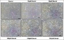
          Studying the viability of cells confirms the IC 50 of Steviol in MCF-7 cells which was 185 μM. The data obtained from fluorescence-activated cell sorter analysis reveal Steviol-mediated G2/M-phase arrest ( P < 0.05) in addition to the presence of evident sub-G0/G1 peak ( P < 0.05) in the MCF-7 cells, signifying the ongoing apoptosis.

          Conclusion:

          Thus, results suggest that induction of apoptosis in MCF-7 cells was due to dose-dependent effect of Steviol. Our first in vitro findings indicate Steviol as a promising candidate for the treatment of breast cancer.

          SUMMARY

          • Steviol remarkably inhibited the growth MCF-7 HBCCs in a dose dependent manner

          • It abolishes cell cycle progression by arresting cells at G2/M phase

          • Steviol induces the cells to undergo apoptosis

          • Steviol induces the cells to generate reactive oxygen species (ROS).

          Abbreviations used: MCF-7: Michigan Cancer Foundation-7; SRB: Sulforhodamine-B assay; FACS: Fluorescence-activated cell sorter; ROS: Reactive oxygen species; DNA: Deoxyribonucleic acid.

          Related collections

          Most cited references23

          • Record: found
          • Abstract: found
          • Article: not found

          Epidermal growth factor (EGF)-induced generation of hydrogen peroxide. Role in EGF receptor-mediated tyrosine phosphorylation.

          Recent evidence indicates that reactive oxygen species (ROS) may function as intracellular messengers in receptor signaling pathways. The possible role of ROS in epidermal growth factor (EGF) signaling was therefore investigated. Stimulation of A431 human epidermoid carcinoma cells with EGF resulted in a transient increase in the intracellular concentration of ROS, measured with the oxidation-sensitive fluorescent probe 2',7'-dichlorofluorescin diacetate and laser-scanning confocal microscopy. The predominant ROS produced appeared to be H2O2, because the EGF-induced increase in fluorescence was completely abolished by incorporation of catalase into the cells by electroporation. The elimination of H2O2 by catalase also inhibited the EGF-induced tyrosine phosphorylation of various cellular proteins including the EGF receptor and phospholipase C-gamma1. The dependence of H2O2 production on the intrinsic tyrosine kinase activity of the EGF receptor and the autophosphorylation sites located in its COOH-terminal tail was investigated. EGF failed to induce H2O2 generation in cells expressing a kinase-inactive EGF receptor. However, normal H2O2 generation was observed in cells expressing a mutant receptor from which the 126 COOH-terminal amino acids had been deleted to remove four (out of the total of five) autophosphorylation sites. These results suggest that EGF-induced H2O2 formation requires the kinase activity but probably not the autophosphorylation sites of the EGF receptor and that inhibition of protein tyrosine phosphatase activity by H2O2 may be required for EGF-induced protein tyrosine phosphorylation to be manifested.
            Bookmark
            • Record: found
            • Abstract: found
            • Article: not found

            Activation of mitogen-activated protein kinase by H2O2. Role in cell survival following oxidant injury.

            The mitogen-activated protein kinase (MAPK) family is comprised of key regulatory proteins that control the cellular response to both proliferation and stress signals. In this study we investigated the factors controlling MAPK activation by H2O2 and explored the impact of altering the pathways to kinase activation on cell survival following H2O2 exposure. Potent activation (10-20-fold) of extracellular signal-regulated protein kinase (ERK2) occurred within 10 min of H2O2 treatment, whereupon rapid inactivation ensued. H2O2 activated ERK2 in several cell types and also moderately activated (3-5-fold) both c-Jun N-terminal kinase and p38/RK/CSBP. Additionally, H2O2 increased the mRNA expression of MAPK-dependent genes c-jun, c-fos, and MAPK phosphatase-1. Suramin pretreatment completely inhibited H2O2 stimulation of ERK2, highlighting a role for growth factor receptors in this activation. Further, ERK2 activation by H2O2 was blocked by pretreatment with either N-acetyl-cysteine, o-phenanthroline, or mannitol, indicating that metal-catalyzed free radical formation mediates the initiation of signal transduction by H2O2. H2O2-stimulated activation of ERK2 was abolished in PC12 cells by inducible or constitutive expression of the dominant negative Ras-N-17 allele. Interestingly, PC12/Ras-N-17 cells were more sensitive than wild-type PC12 cells to H2O2 toxicity. Moreover, NIH 3T3 cells expressing constitutively active MAPK kinase (MEK, the immediate upstream regulator of ERK) were more resistant to H2O2 toxicity, while those expressing kinase-defective MEK were more sensitive, than cells expressing wild-type MEK. Taken together, these studies provide insight into mechanisms of MAPK regulation by H2O2 and suggest that ERK plays a critical role in cell survival following oxidant injury.
              Bookmark
              • Record: found
              • Abstract: found
              • Article: not found

              Stevioside and related compounds: therapeutic benefits beyond sweetness.

              Stevioside, an abundant component of Stevia rebaudiana leaf, has become well-known for its intense sweetness (250-300 times sweeter than sucrose) and is used as a non-caloric sweetener in several countries. A number of studies have suggested that, beside sweetness, stevioside along with related compounds, which include rebaudioside A (second most abundant component of S. rebaudiana leaf), steviol and isosteviol (metabolic components of stevioside) may also offer therapeutic benefits, as they have anti-hyperglycemic, anti-hypertensive, anti-inflammatory, anti-tumor, anti-diarrheal, diuretic, and immunomodulatory actions. It is of interest to note that their effects on plasma glucose level and blood pressure are only observed when these parameters are higher than normal. As steviol can interact with drug transporters, its role as a drug modulator is proposed. This review summarizes the current knowledge of the pharmacological actions, therapeutic applications, pharmacokinetics and safety of stevioside and related compounds. Although much progress has been made concerning their biological and pharmacological effects, questions regarding chemical purity and safety remain unsolved. These issues are discussed to help guide future research directions.
                Bookmark

                Author and article information

                Journal
                Pharmacogn Mag
                Pharmacogn Mag
                PM
                Pharmacognosy Magazine
                Medknow Publications & Media Pvt Ltd (India )
                0973-1296
                0976-4062
                Jul-Sep 2017
                19 July 2017
                : 13
                : 51
                : 345-350
                Affiliations
                [1]Centre of Biotechnology, University of Allahabad, Allahabad, Uttar Pradesh, India
                [1 ]Division of Biochemistry, Central Drug Research Institute, Lucknow, Uttar Pradesh, India
                Author notes
                Correspondence: Dr. Ena Gupta, Centre of Biotechnology, University of Allahabad, Allahabad, Uttar Pradesh, India. E-mail: enaravish@ 123456gmail.com
                Article
                PM-13-345
                10.4103/pm.pm_29_17
                5551348
                1d72acb7-e38f-4081-a3e4-e70030f247a4
                Copyright: © 2017 Pharmacognosy Magazine

                This is an open access article distributed under the terms of the Creative Commons Attribution-NonCommercial-ShareAlike 3.0 License, which allows others to remix, tweak, and build upon the work non-commercially, as long as the author is credited and the new creations are licensed under the identical terms.

                History
                : 26 January 2017
                : 21 February 2017
                Categories
                Original Article

                Pharmacology & Pharmaceutical medicine
                apoptosis,michigan cancer foundation-7,stevia rebaudiana,steviol

                Comments

                Comment on this article