20
views
0
recommends
+1 Recommend
0 collections
    0
    shares
      • Record: found
      • Abstract: found
      • Article: found
      Is Open Access

      Bakers' cyst and tibiofemoral abnormalities are more distinctive MRI features of symptomatic osteoarthritis than patellofemoral abnormalities

      research-article

      Read this article at

      Bookmark
          There is no author summary for this article yet. Authors can add summaries to their articles on ScienceOpen to make them more accessible to a non-specialist audience.

          Abstract

          Objective

          To investigate which structural MR abnormalities discriminate symptomatic knee osteoarthritis (OA), taking co-occurrence of abnormalities in all compartments into account.

          Methods

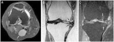
          The Netherlands Epidemiology of Obesity (NEO) study is a population-based cohort aged 45–65 years. In 1285 participants (median age 56 years, 55% women, median body mass index (BMI) 30 kg/m 2), MRI of the right knee were obtained. Structural abnormalities (osteophytes, cartilage loss, bone marrow lesions (BMLs), subchondral cysts, meniscal abnormalities, effusion, Baker's cyst) at 9 patellofemoral and tibiofemoral locations were scored following the knee OA scoring system. Symptomatic OA in the imaged knee was defined following the American College of Rheumatology criteria. Logistic ridge regression analyses were used to investigate which structural abnormalities discriminate best between individuals with and without symptomatic OA, crude and adjusted for age, sex and BMI.

          Results

          Symptomatic knee OA was present in 177 individuals. Structural MR abnormalities were highly frequent both in individuals with OA and in those without. Baker's cysts showed the highest adjusted regression coefficient (0.293) for presence of symptomatic OA, followed by osteophytes and BMLs in the medial tibiofemoral compartment (0.185–0.279), osteophytes in the medial trochlear facet (0.262) and effusion (0.197).

          Conclusions

          Baker's cysts discriminate best between individuals with and without symptomatic knee OA. Structural MR abnormalities, especially in the medial side of the tibiofemoral joint and effusion, add further in discriminating symptomatic OA. Baker's cysts may present as a target for treatment.

          Related collections

          Most cited references23

          • Record: found
          • Abstract: found
          • Article: not found

          The association of bone marrow lesions with pain in knee osteoarthritis.

          The cause of pain in osteoarthritis is unknown. Bone has pain fibers, and marrow lesions, which are thought to represent edema, have been noted in osteoarthritis. To determine whether bone marrow lesions on magnetic resonance imaging (MRI) are associated with pain in knee osteoarthritis. Cross-sectional observational study. Veterans Affairs Medical Center. 401 persons (mean age, 66.8 years) with knee osteoarthritis on radiography who were drawn from clinics in the Veterans Administration health care system and from the community. Of these persons, 351 had knee pain and 50 had no knee pain. Knee radiography and MRI of one knee were performed in all participants. Those with knee pain quantified the severity of their pain. On MRI, coronal T(2)-weighted fat-saturated images were used to score the size of bone marrow lesions, and each knee was characterized as having any lesion or any large lesion. The prevalence of lesions and large lesions in persons with and without knee pain was compared; in participants with knee pain, the presence of lesions was correlated with severity of pain. Bone marrow lesions were found in 272 of 351 (77.5%) persons with painful knees compared with 15 of 50 (30%) persons with no knee pain (P < 0.001). Large lesions were present almost exclusively in persons with knee pain (35.9% vs. 2%; P < 0.001). After adjustment for severity of radiographic disease, effusion, age, and sex, lesions and large lesions remained associated with the occurrence of knee pain. Among persons with knee pain, bone marrow lesions were not associated with pain severity. Bone marrow lesions on MRI are strongly associated with the presence of pain in knee osteoarthritis.
            Bookmark
            • Record: found
            • Abstract: found
            • Article: not found

            Osteoarthritis of the knee: association between clinical features and MR imaging findings.

            To prospectively evaluate the association between clinical features and structural abnormalities found at magnetic resonance (MR) imaging in patients with osteoarthritis (OA) of the knee. The study was approved by the institutional medical ethics review board. Written informed consent was obtained from each patient. MR images of the knee were obtained from 205 (42 [20%] men, 163 [80%] women; median age, 60 years; range, 43-77 years) patients in whom symptomatic OA at multiple joint sites was diagnosed. MR images were analyzed for various abnormalities of OA. All patients were interviewed concerning pain and stiffness in the knee that was imaged. Odds ratios (ORs) with 99% confidence intervals (CIs) were used to determine the association between the imaging findings and clinical features of OA. A large joint effusion was associated with pain (OR, 9.99; 99% CI: 1.28, 149) and stiffness (OR, 4.67; 99% CI: 1.26, 26.1). The presence of an osteophyte in the patellofemoral compartment (OR, 2.25; 99% CI: 1.06, 4.77) was associated with pain. All other imaging findings, including focal or diffuse cartilaginous abnormalities, subchondral cysts, bone marrow edema, subluxation of the meniscus, meniscal tears, or Baker cysts, were not associated with symptoms. Findings of this study indicate that only two associations exist between clinical symptoms and structural findings found on MR images in patients with OA of the knee. Copyright (c) RSNA, 2006.
              Bookmark
              • Record: found
              • Abstract: found
              • Article: not found

              Receiver operating characteristic (ROC) methodology: the state of the art.

              Receiver operating characteristic (ROC) methodology has been increasingly used in medical applications in the last 10 years. The text by Swets and Pickett has popularized the technique and the journal Medical Decision Making (1981--) provides a forum for further methodologic issues. In this article, I will (1) describe the nature of the data generated by ROC studies; (2) evaluate the choices of summary indices of performance (accuracy); (3) outline the data-analytic techniques used, and how to incorporate data from multiple observers and multiple "readings"; (4) review proposed alternatives to the commonly used binormal ROC model; and (5) discuss issues, such as verification bias, and challenges, such as multicenter comparative imaging studies and the difficulty of obtaining "truth data", which need to be addressed when adapting ROC methods to medical contexts.
                Bookmark

                Author and article information

                Journal
                RMD Open
                RMD Open
                rmdopen
                rmdopen
                RMD Open
                BMJ Publishing Group (BMA House, Tavistock Square, London, WC1H 9JR )
                2056-5933
                2016
                17 May 2016
                : 2
                : 1
                : e000234
                Affiliations
                [1 ]Department of Rheumatology, Leiden University Medical Center , Leiden, The Netherlands
                [2 ]Department of Medical Statistics and Bio-informatics,Leiden University Medical Center , Leiden, The Netherlands
                [3 ]Department of Radiology, Leiden University Medical Center , Leiden, The Netherlands
                [4 ]Department of Clinical Epidemiology,Leiden University Medical Center , Leiden, The Netherlands
                [5 ]Department of Thrombosis and Homeostasis, Leiden University Medical Center , Leiden, The Netherlands
                Author notes
                [Correspondence to ] Dr A W Visser; a.w.visser@ 123456lumc.nl
                Article
                rmdopen-2015-000234
                10.1136/rmdopen-2015-000234
                4879339
                27252896
                1e40b549-1551-4e6e-be78-79bb5238d027
                Published by the BMJ Publishing Group Limited. For permission to use (where not already granted under a licence) please go to http://www.bmj.com/company/products-services/rights-and-licensing/

                This is an Open Access article distributed in accordance with the Creative Commons Attribution Non Commercial (CC BY-NC 4.0) license, which permits others to distribute, remix, adapt, build upon this work non-commercially, and license their derivative works on different terms, provided the original work is properly cited and the use is non-commercial. See: http://creativecommons.org/licenses/by-nc/4.0/

                History
                : 17 December 2015
                : 11 April 2016
                : 14 April 2016
                Funding
                Funded by: Reumafonds, http://dx.doi.org/10.13039/501100006315;
                Award ID: 10-1-309
                Categories
                Osteoarthritis
                1506
                Original article

                epidemiology,knee osteoarthritis,magnetic resonance imaging

                Comments

                Comment on this article