22
views
0
recommends
+1 Recommend
0 collections
    0
    shares
      • Record: found
      • Abstract: found
      • Article: not found

      Apertureless scanning near-field optical microscopy of sparsely labeled tobacco mosaic viruses and the intermediate filament desmin

      research-article

      Read this article at

      ScienceOpenPublisherPMC
      Bookmark
          There is no author summary for this article yet. Authors can add summaries to their articles on ScienceOpen to make them more accessible to a non-specialist audience.

          Summary

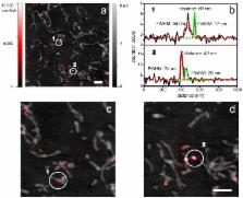
          Both fluorescence imaging and atomic force microscopy (AFM) are highly versatile and extensively used in applications ranging from nanotechnology to life sciences. In fluorescence microscopy luminescent dyes serve as position markers. Moreover, they can be used as active reporters of their local vicinity. The dipolar coupling of the tip with the incident light and the fluorophore give rise to a local field and fluorescence enhancement. AFM topographic imaging allows for resolutions down to the atomic scale. It can be operated in vacuum, under ambient conditions and in liquids. This makes it ideal for the investigation of a wide range of different samples. Furthermore an illuminated AFM cantilever tip apex exposes strongly confined non-propagating electromagnetic fields that can serve as a coupling agent for single dye molecules. Thus, combining both techniques by means of apertureless scanning near-field optical microscopy (aSNOM) enables concurrent high resolution topography and fluorescence imaging. Commonly, among the various (apertureless) SNOM approaches metallic or metallized probes are used. Here, we report on our custom-built aSNOM setup, which uses commercially available monolithic silicon AFM cantilevers. The field enhancement confined to the tip apex facilitates an optical resolution down to 20 nm. Furthermore, the use of standard mass-produced AFM cantilevers spares elaborate probe production or modification processes. We investigated tobacco mosaic viruses and the intermediate filament protein desmin. Both are mixed complexes of building blocks, which are fluorescently labeled to a low degree. The simultaneous recording of topography and fluorescence data allows for the exact localization of distinct building blocks within the superordinate structures.

          Abstract

          Related collections

          Most cited references32

          • Record: found
          • Abstract: not found
          • Article: not found

          Optical stethoscopy: Image recording with resolution λ/20

            Bookmark
            • Record: found
            • Abstract: found
            • Article: not found

            Radiative decay engineering: biophysical and biomedical applications.

            Fluorescence spectroscopy is a widely used research tool in biochemistry and molecular biology. Fluorescence has also become the dominant method enabling the revolution in medical diagnostics, DNA sequencing, and genomics. To date all the fluorescence observables, including spectral shifts, anisotropies, quantum yields, and lifetimes, have all been utilized in basic and applied uses of fluorescence. In this forward-looking article we describe a new opportunity in fluorescence, radiative decay engineering (RDE). By RDE we mean modifying the emission of fluorophores or chromophores by increasing or decreasing their radiative decay rates. In most fluorescence experiments the radiative rates are not changed because these rates depend on the extinction coefficient of the fluorophore. This intrinsic rate is not changed by quenching and is only weakly dependent on environmental effects. Spectral changes are usually caused by changes in the nonradiative rates resulting from quenching or resonance energy transfer. These processes affect the emission by providing additional routes for decay of the excited states without emission. In contrast to the relatively constant radiative rates in free solution, it is known that the radiative rates can be modified by placing the fluorophores at suitable distances from metallic surfaces and particles. This Review summarizes results from the physics literature which demonstrate the effects of metallic surfaces, colloids, or islands on increasing or decreasing emissive rates, increasing the quantum yields of low quantum yield chromophores, decreasing the lifetimes, and directing the typically isotropic emission in specific directions. These effects are not due to reflection of the emitted photons, but rather as the result of the fluorophore dipole interacting with free electrons in the metal. These interactions change the intensity and temporal and spatial distribution of the radiation. We describe the unusual effects expected from increases in the radiative rates with reference to intrinsic and extrinsic biochemical fluorophores. For instance, the decreased lifetime can result in an effective increase in photostability. Proximity to nearby metallic surfaces can also increase the local field and modify the rate of excitation. We predict that the appropriate localization of fluorophores near particles can result in usefully high emission from "nonfluorescent" molecules and million-fold increases in the number of photons observable from each fluorophore. We also describe how RDE can be applied to medical testing and biotechnology. As one example we predict that nearby metal surfaces can be used to increase the low intrinsic quantum yields of nucleic acids and make unlabeled DNA detectable using its intrinsic metal-enhanced fluorescence. Copyright 2001 Academic Press.
              Bookmark
              • Record: found
              • Abstract: found
              • Article: not found

              Enhancement and quenching of single-molecule fluorescence.

              We present an experimental and theoretical study of the fluorescence rate of a single molecule as a function of its distance to a laser-irradiated gold nanoparticle. The local field enhancement leads to an increased excitation rate whereas nonradiative energy transfer to the particle leads to a decrease of the quantum yield (quenching). Because of these competing effects, previous experiments showed either fluorescence enhancement or fluorescence quenching. By varying the distance between molecule and particle we show the first experimental measurement demonstrating the continuous transition from fluorescence enhancement to fluorescence quenching. This transition cannot be explained by treating the particle as a polarizable sphere in the dipole approximation.
                Bookmark

                Author and article information

                Contributors
                Role: Guest Editor
                Role: Guest Editor
                Journal
                Beilstein J Nanotechnol
                Beilstein J Nanotechnol
                Beilstein Journal of Nanotechnology
                Beilstein-Institut (Trakehner Str. 7-9, 60487 Frankfurt am Main, Germany )
                2190-4286
                2013
                11 September 2013
                : 4
                : 510-516
                Affiliations
                [1 ]Experimental Biophysics and Applied Nanoscience, Faculty of Physics and Bielefeld Institute for Biophysics and Nanoscience (BINAS), Bielefeld University, Universitätsstrasse 25, D-33615 Bielefeld, Germany
                [2 ]Department of Molecular Biology and Plant Virology, Institute of Biology, University of Stuttgart, Pfaffenwaldring 57, D-70569 Stuttgart, Germany
                [3 ]Erich and Hanna Klessmann Institute for Cardiovascular Research & Development (EHKI), Heart and Diabetes Center NRW, Ruhr University Bochum, Georgstraße 11, D-32545 Bad Oeynhausen, Germany
                [4 ]Libin Cardiovascular Institute of Alberta, Department of Cardiac Sciences, University of Calgary, 3280 Hospital Drive NW, T2N4Z6, AB, Canada
                Article
                10.3762/bjnano.4.60
                3778390
                24062977
                1fcc5c61-9015-4f9e-9332-c86fde258946
                Copyright © 2013, Harder et al; licensee Beilstein-Institut.

                This is an Open Access article under the terms of the Creative Commons Attribution License ( http://creativecommons.org/licenses/by/2.0), which permits unrestricted use, distribution, and reproduction in any medium, provided the original work is properly cited.

                The license is subject to the Beilstein Journal of Nanotechnology terms and conditions: ( http://www.beilstein-journals.org/bjnano)

                History
                : 18 April 2013
                : 22 August 2013
                Categories
                Full Research Paper
                Nanoscience
                Nanotechnology

                apertureless scanning near-field optical microscope,atomic force microscopy,fluorescence microscopy

                Comments

                Comment on this article