20
views
0
recommends
+1 Recommend
0 collections
    0
    shares
      • Record: found
      • Abstract: found
      • Article: found
      Is Open Access

      Oligodendrocyte Precursor Cells Modulate the Neuronal Network by Activity-Dependent Ectodomain Cleavage of Glial NG2

      research-article

      Read this article at

      Bookmark
          There is no author summary for this article yet. Authors can add summaries to their articles on ScienceOpen to make them more accessible to a non-specialist audience.

          Abstract

          This study shows that the activity of neurons can trigger shedding of a protein, NG2, from the surface of oligodendrocyte precursor cells; this protein in turn modulates synaptic transmission, revealing a two-way conversation between neurons and glia.

          Abstract

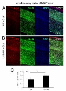
          The role of glia in modulating neuronal network activity is an important question. Oligodendrocyte precursor cells (OPC) characteristically express the transmembrane proteoglycan nerve-glia antigen 2 (NG2) and are unique glial cells receiving synaptic input from neurons. The development of NG2+ OPC into myelinating oligodendrocytes has been well studied, yet the retention of a large population of synapse-bearing OPC in the adult brain poses the question as to additional functional roles of OPC in the neuronal network. Here we report that activity-dependent processing of NG2 by OPC-expressed secretases functionally regulates the neuronal network. NG2 cleavage by the α-secretase ADAM10 yields an ectodomain present in the extracellular matrix and a C-terminal fragment that is subsequently further processed by the γ-secretase to release an intracellular domain. ADAM10-dependent NG2 ectodomain cleavage and release (shedding) in acute brain slices or isolated OPC is increased by distinct activity-increasing stimuli. Lack of NG2 expression in OPC (NG2-knockout mice), or pharmacological inhibition of NG2 ectodomain shedding in wild-type OPC, results in a striking reduction of N-methyl-D-aspartate (NMDA) receptor-dependent long-term potentiation (LTP) in pyramidal neurons of the somatosensory cortex and alterations in the subunit composition of their α-amino-3-hydroxy-5-methyl-4-isoxazolepr opionicacid (AMPA) receptors. In NG2-knockout mice these neurons exhibit diminished AMPA and NMDA receptor-dependent current amplitudes; strikingly AMPA receptor currents can be rescued by application of conserved LNS protein domains of the NG2 ectodomain. Furthermore, NG2-knockout mice exhibit altered behavior in tests measuring sensorimotor function. These results demonstrate for the first time a bidirectional cross-talk between OPC and the surrounding neuronal network and demonstrate a novel physiological role for OPC in regulating information processing at neuronal synapses.

          Author Summary

          Although glial cells substantially outnumber neurons in the mammalian brain, much remains to be discovered regarding their functions. Among glial cells, oligodendrocyte precursors differentiate into oligodendrocytes, whose function is to enwrap nerves with myelin to ensure proper impulse conduction. However, oligodendrocyte precursors also comprise a stable population in all major regions of the adult brain, making up around 5% of the total number of neurons and glia. Synapses are classically formed between neurons. Nonetheless, oligodendrocyte precursors are unique among glial cells in that they receive direct synaptic input from different types of neurons; whether OPC also send signals to neurons is still unknown. Here we show a bidirectional communication between neurons and oligodendrocyte precursors: neuronal activity regulates the cleavage of a glial membrane protein and the release of an extracellular domain that in turn modulates synaptic transmission between neurons. Our data thus show that a particular subtype of glial cells, oligodendrocyte precursors, functionally integrate into the neuronal network and we link this bidirectional signaling to mouse behavior and disease.

          Related collections

          Most cited references79

          • Record: found
          • Abstract: found
          • Article: not found

          NG2-expressing glial progenitor cells: an abundant and widespread population of cycling cells in the adult rat CNS.

          M. Dawson (2003)
          Glial progenitor cells of the developing CNS committed to the oligodendrocyte lineage (OPCs) express the chondroitin sulfate proteoglycan, NG2. A proportion of OPCs fail to differentiate past the stage at which they express NG2 and the lipid antigen O4 and persist in the adult CNS in a phenotypically immature form. However, the physiological function of NG2(+) cells in the adult CNS is unknown. Using antibodies against NG2 we show that NG2 is expressed by a distinct cell population in the mature CNS with the homogeneous antigenic phenotype of oligodendrocyte progenitors. The morphology of NG2(+) OPCs varies from region to region, reflecting the different structural environments, but they appear to represent a homogeneous population within any one gray or white matter region. A study of nine CNS regions showed that NG2(+) OPCs are numerous throughout the CNS and numbers in the white matter are only 1.5 times that in the gray. Whereas the ratio of OPCs to myelinating oligodendrocytes in the spinal cord gray and white matter approximates 1:4, gray matter regions of the forebrain have a 1:1 ratio, a phenomenon that will have consequences for oligodendrocyte replacement following demyelination. BrdU incorporation experiments showed that NG2(+) cells are the major dividing cell population of the adult rat CNS. Since very little apoptosis was detected and BrdU became increasingly present in oligodendrocytes after a 10-day pulse chase, with a concomitant decrease in NG2(+) BrdU incorporating cells, we suggest that the size of the oligodendrocyte population may actually increase during adult life.
            Bookmark
            • Record: found
            • Abstract: found
            • Article: not found

            Glutamatergic synapses on oligodendrocyte precursor cells in the hippocampus.

            Fast excitatory neurotransmission in the central nervous system occurs at specialized synaptic junctions between neurons, where a high concentration of glutamate directly activates receptor channels. Low-affinity AMPA (alpha-amino-3-hydroxy-5-methyl isoxazole propionic acid) and kainate glutamate receptors are also expressed by some glial cells, including oligodendrocyte precursor cells (OPCs). However, the conditions that result in activation of glutamate receptors on these non-neuronal cells are not known. Here we report that stimulation of excitatory axons in the hippocampus elicits inward currents in OPCs that are mediated by AMPA receptors. The quantal nature of these responses and their rapid kinetics indicate that they are produced by the exocytosis of vesicles filled with glutamate directly opposite these receptors. Some of these AMPA receptors are permeable to calcium ions, providing a link between axonal activity and internal calcium levels in OPCs. Electron microscopic analysis revealed that vesicle-filled axon terminals make synaptic junctions with the processes of OPCs in both the young and adult hippocampus. These results demonstrate the existence of a rapid signalling pathway from pyramidal neurons to OPCs in the mammalian hippocampus that is mediated by excitatory, glutamatergic synapses.
              Bookmark
              • Record: found
              • Abstract: found
              • Article: not found

              Polydendrocytes (NG2 cells): multifunctional cells with lineage plasticity.

              NG2 cells (also known as polydendrocytes) are a population of CNS cells that are distinct from neurons, mature oligodendrocytes, astrocytes and microglia. They can be identified by the expression of the proteoglycan NG2, have a highly branched morphology and are distributed throughout the grey and white matter. They differentiate into oligodendrocytes in vitro and have often been equated with oligodendrocyte precursor cells. However, whether polydendrocytes are multipotential cells that can give rise to neurons and astrocytes as well as oligodendrocytes is now highly debated. Furthermore, electrophysiological studies indicate that polydendrocytes receive synaptic input from neurons, suggesting that they are integrated in the neural network. This Review highlights recent findings and unresolved questions related to the lineage and function of polydendrocytes in the CNS.
                Bookmark

                Author and article information

                Contributors
                Role: Academic Editor
                Journal
                PLoS Biol
                PLoS Biol
                plos
                plosbiol
                PLoS Biology
                Public Library of Science (San Francisco, USA )
                1544-9173
                1545-7885
                November 2014
                11 November 2014
                : 12
                : 11
                : e1001993
                Affiliations
                [1 ]Molecular Cell Biology, Department of Biology, Johannes Gutenberg University Mainz, Mainz, Germany
                [2 ]Institute of Physiology, University Medical Center of the Johannes-Gutenberg University Mainz, Mainz, Germany
                [3 ]Leibniz Institute for Neurobiology, Magdeburg, Germany
                [4 ]Department of Psychiatry and Psychotherapy, University Medicine Mainz, Mainz, Germany
                [5 ]Institute of Physiological Chemistry, University Medical Center Mainz, Mainz, Germany
                [6 ]Mouse Behavior Outcome Unit, Focus Program Translational Neurosciences (FTN), Johannes Gutenberg University Mainz, Mainz, Germany
                Stanford University School of Medicine, United States of America
                Author notes

                The authors have declared that no competing interests exist.

                The author(s) have made the following declarations about their contributions: Conceived and designed the experiments: DS AN RF KR BL TM JT. Performed the experiments: DS AN JS DM KR. Analyzed the data: DS AN JS KR JT. Contributed reagents/materials/analysis tools: FB KE SP. Wrote the paper: DS AN JT. Contributed to the writing of the manuscript: RF TM BL KR KE.

                [¤]

                Current address: Department of Clinical Neurobiology, University Hospital and German Cancer Center (DKFZ) Heidelberg, Heidelberg, Germany

                ¶ JT and TM also contributed equally to this work.

                Article
                PBIOLOGY-D-14-00151
                10.1371/journal.pbio.1001993
                4227637
                25387269
                25a9b815-3186-4880-a2d3-0b5fa7828b99
                Copyright @ 2014

                This is an open-access article distributed under the terms of the Creative Commons Attribution License, which permits unrestricted use, distribution, and reproduction in any medium, provided the original author and source are credited.

                History
                : 14 January 2014
                : 29 September 2014
                Page count
                Pages: 17
                Funding
                This work was financed by the Deutsche Forschungsgemeinschaft (DFG) (TR 1008/1-1, CRC TRR 128 TP B07, SPP 1172 TR 231 and GRK1044/2) to JT, the DFG (CRC 8080, TP A7) to TM, and the Schram Stiftung (T287/21796/2011) to RF. The funders had no role in study design, data collection and analysis, decision to publish, or preparation of the manuscript.
                Categories
                Research Article
                Biology and Life Sciences
                Neuroscience
                Behavioral Neuroscience
                Cellular Neuroscience
                Molecular Neuroscience
                Neural Networks
                Research and Analysis Methods
                Specimen Preparation and Treatment
                Mechanical Treatment of Specimens
                Specimen Disruption
                Electroporation

                Life sciences
                Life sciences

                Comments

                Comment on this article