14
views
0
recommends
+1 Recommend
0 collections
    0
    shares
      • Record: found
      • Abstract: found
      • Article: found
      Is Open Access

      The Anti-Inflammatory and Antioxidant Properties of n-3 PUFAs: Their Role in Cardiovascular Protection

      review-article

      Read this article at

      Bookmark
          There is no author summary for this article yet. Authors can add summaries to their articles on ScienceOpen to make them more accessible to a non-specialist audience.

          Abstract

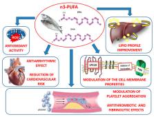
          Polyunsaturated fatty acids (n-3 PUFAs) are long-chain polyunsaturated fatty acids with 18, 20 or 22 carbon atoms, which have been found able to counteract cardiovascular diseases. Eicosapentaenoic acid (EPA) and docosahexaenoic acid (DHA), in particular, have been found to produce both vaso- and cardio-protective response via modulation of membrane phospholipids thereby improving cardiac mitochondrial functions and energy production. However, antioxidant properties of n-3 PUFAs, along with their anti-inflammatory effect in both blood vessels and cardiac cells, seem to exert beneficial effects in cardiovascular impairment. In fact, dietary supplementation with n-3 PUFAs has been demonstrated to reduce oxidative stress-related mitochondrial dysfunction and endothelial cell apoptosis, an effect occurring via an increased activity of endogenous antioxidant enzymes. On the other hand, n-3 PUFAs have been shown to counteract the release of pro-inflammatory cytokines in both vascular tissues and in the myocardium, thereby restoring vascular reactivity and myocardial performance. Here we summarize the molecular mechanisms underlying the anti-oxidant and anti-inflammatory effect of n-3 PUFAs in vascular and cardiac tissues and their implication in the prevention and treatment of cardiovascular disease.

          Related collections

          Most cited references108

          • Record: found
          • Abstract: found
          • Article: found
          Is Open Access

          Reperfusion injury and reactive oxygen species: The evolution of a concept☆

          Reperfusion injury, the paradoxical tissue response that is manifested by blood flow-deprived and oxygen-starved organs following the restoration of blood flow and tissue oxygenation, has been a focus of basic and clinical research for over 4-decades. While a variety of molecular mechanisms have been proposed to explain this phenomenon, excess production of reactive oxygen species (ROS) continues to receive much attention as a critical factor in the genesis of reperfusion injury. As a consequence, considerable effort has been devoted to identifying the dominant cellular and enzymatic sources of excess ROS production following ischemia-reperfusion (I/R). Of the potential ROS sources described to date, xanthine oxidase, NADPH oxidase (Nox), mitochondria, and uncoupled nitric oxide synthase have gained a status as the most likely contributors to reperfusion-induced oxidative stress and represent priority targets for therapeutic intervention against reperfusion-induced organ dysfunction and tissue damage. Although all four enzymatic sources are present in most tissues and are likely to play some role in reperfusion injury, priority and emphasis has been given to specific ROS sources that are enriched in certain tissues, such as xanthine oxidase in the gastrointestinal tract and mitochondria in the metabolically active heart and brain. The possibility that multiple ROS sources contribute to reperfusion injury in most tissues is supported by evidence demonstrating that redox-signaling enables ROS produced by one enzymatic source (e.g., Nox) to activate and enhance ROS production by a second source (e.g., mitochondria). This review provides a synopsis of the evidence implicating ROS in reperfusion injury, the clinical implications of this phenomenon, and summarizes current understanding of the four most frequently invoked enzymatic sources of ROS production in post-ischemic tissue.
            Bookmark
            • Record: found
            • Abstract: found
            • Article: not found

            Vascular Smooth Muscle Cells and Arterial Stiffening: Relevance in Development, Aging, and Disease.

            The cushioning function of large arteries encompasses distension during systole and recoil during diastole which transforms pulsatile flow into a steady flow in the microcirculation. Arterial stiffness, the inverse of distensibility, has been implicated in various etiologies of chronic common and monogenic cardiovascular diseases and is a major cause of morbidity and mortality globally. The first components that contribute to arterial stiffening are extracellular matrix (ECM) proteins that support the mechanical load, while the second important components are vascular smooth muscle cells (VSMCs), which not only regulate actomyosin interactions for contraction but mediate also mechanotransduction in cell-ECM homeostasis. Eventually, VSMC plasticity and signaling in both conductance and resistance arteries are highly relevant to the physiology of normal and early vascular aging. This review summarizes current concepts of central pressure and tensile pulsatile circumferential stress as key mechanical determinants of arterial wall remodeling, cell-ECM interactions depending mainly on the architecture of cytoskeletal proteins and focal adhesion, the large/small arteries cross-talk that gives rise to target organ damage, and inflammatory pathways leading to calcification or atherosclerosis. We further speculate on the contribution of cellular stiffness along the arterial tree to vascular wall stiffness. In addition, this review provides the latest advances in the identification of gene variants affecting arterial stiffening. Now that important hemodynamic and molecular mechanisms of arterial stiffness have been elucidated, and the complex interplay between ECM, cells, and sensors identified, further research should study their potential to halt or to reverse the development of arterial stiffness.
              Bookmark
              • Record: found
              • Abstract: found
              • Article: not found

              Impact of Oxidative Stress on the Heart and Vasculature: Part 2 of a 3-Part Series.

              Vascular disease and heart failure impart an enormous burden in terms of global morbidity and mortality. Although there are many different causes of cardiac and vascular disease, most causes share an important pathological mechanism: oxidative stress. In the failing heart, oxidative stress occurs in the myocardium and correlates with left ventricular dysfunction. Reactive oxygen species (ROS) negatively affect myocardial calcium handling, cause arrhythmia, and contribute to cardiac remodeling by inducing hypertrophic signaling, apoptosis, and necrosis. Similarly, oxidative balance in the vasculature is tightly regulated by a wealth of pro- and antioxidant systems that orchestrate region-specific ROS production and removal. Reactive oxygen species also regulate multiple vascular cell functions, including endothelial and smooth muscle cell growth, proliferation, and migration; angiogenesis; apoptosis; vascular tone; host defenses; and genomic stability. However, excessive levels of ROS promote vascular disease through direct and irreversible oxidative damage to macromolecules, as well as disruption of redox-dependent vascular wall signaling processes.
                Bookmark

                Author and article information

                Journal
                Biomedicines
                Biomedicines
                biomedicines
                Biomedicines
                MDPI
                2227-9059
                25 August 2020
                September 2020
                : 8
                : 9
                : 306
                Affiliations
                [1 ]Institute of Research for Food Safety and Health (IRC-FSH), Department of Health Sciences, University “Magna Graecia” of Catanzaro, 88100 Catanzaro, Italy; oppedisanof@ 123456libero.it (F.O.); robertamacri85@ 123456gmail.com (R.M.); gliozzi@ 123456unicz.it (M.G.); v.musolino@ 123456unicz.it (V.M.); carresi@ 123456unicz.it (C.C.); jessicamaiuolo@ 123456virgilio.it (J.M.); boscofrancescabf@ 123456libero.it (F.B.); saverio.nucera@ 123456hotmail.it (S.N.); mariacaterina.zito@ 123456libero.it (M.C.Z.); lorenzacz808@ 123456gmail.com (L.G.); federicascar87@ 123456gmail.com (F.S.); caterina.nicita@ 123456gmail.com (C.N.); annarita.coppoletta@ 123456libero.it (A.R.C.); rugast1@ 123456gmail.com (S.R.); miriam.scicchitano@ 123456hotmail.it (M.S.); rocco.mollace@ 123456gmail.com (R.M.); palma@ 123456unicz.it (E.P.)
                [2 ]Division of Cardiology, University Hospital Policlinico Tor Vergata, 00133 Rome, Italy
                [3 ]IRCCS San Raffaele Pisana, 00163 Roma, Italy
                Author notes
                [* ]Correspondence: mollace@ 123456unicz.it
                Author information
                https://orcid.org/0000-0003-0930-8987
                https://orcid.org/0000-0003-4199-207X
                Article
                biomedicines-08-00306
                10.3390/biomedicines8090306
                7554783
                32854210
                26596aaa-2ee4-4a9a-aea4-e8ac8f936394
                © 2020 by the authors.

                Licensee MDPI, Basel, Switzerland. This article is an open access article distributed under the terms and conditions of the Creative Commons Attribution (CC BY) license ( http://creativecommons.org/licenses/by/4.0/).

                History
                : 25 June 2020
                : 24 August 2020
                Categories
                Review

                n-3 pufas,oxidative stress,endogenous antioxidants,anti-inflammatory response,cardiovascular diseases

                Comments

                Comment on this article