52
views
0
recommends
+1 Recommend
0 collections
    0
    shares
      • Record: found
      • Abstract: found
      • Article: found
      Is Open Access

      3D tumor spheroid models for in vitro therapeutic screening: a systematic approach to enhance the biological relevance of data obtained

      research-article

      Read this article at

      Bookmark
          There is no author summary for this article yet. Authors can add summaries to their articles on ScienceOpen to make them more accessible to a non-specialist audience.

          Abstract

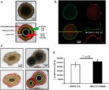
          The potential of a spheroid tumor model composed of cells in different proliferative and metabolic states for the development of new anticancer strategies has been amply demonstrated. However, there is little or no information in the literature on the problems of reproducibility of data originating from experiments using 3D models. Our analyses, carried out using a novel open source software capable of performing an automatic image analysis of 3D tumor colonies, showed that a number of morphology parameters affect the response of large spheroids to treatment. In particular, we found that both spheroid volume and shape may be a source of variability. We also compared some commercially available viability assays specifically designed for 3D models. In conclusion, our data indicate the need for a pre-selection of tumor spheroids of homogeneous volume and shape to reduce data variability to a minimum before use in a cytotoxicity test. In addition, we identified and validated a cytotoxicity test capable of providing meaningful data on the damage induced in large tumor spheroids of up to diameter in 650 μm by different kinds of treatments.

          Related collections

          Most cited references43

          • Record: found
          • Abstract: found
          • Article: not found

          Deconstructing the third dimension: how 3D culture microenvironments alter cellular cues.

          Much of our understanding of the biological mechanisms that underlie cellular functions, such as migration, differentiation and force-sensing has been garnered from studying cells cultured on two-dimensional (2D) glass or plastic surfaces. However, more recently the cell biology field has come to appreciate the dissimilarity between these flat surfaces and the topographically complex, three-dimensional (3D) extracellular environments in which cells routinely operate in vivo. This has spurred substantial efforts towards the development of in vitro 3D biomimetic environments and has encouraged much cross-disciplinary work among biologists, material scientists and tissue engineers. As we move towards more-physiological culture systems for studying fundamental cellular processes, it is crucial to define exactly which factors are operative in 3D microenvironments. Thus, the focus of this Commentary will be on identifying and describing the fundamental features of 3D cell culture systems that influence cell structure, adhesion, mechanotransduction and signaling in response to soluble factors, which - in turn - regulate overall cellular function in ways that depart dramatically from traditional 2D culture formats. Additionally, we will describe experimental scenarios in which 3D culture is particularly relevant, highlight recent advances in materials engineering for studying cell biology, and discuss examples where studying cells in a 3D context provided insights that would not have been observed in traditional 2D systems.
            Bookmark
            • Record: found
            • Abstract: found
            • Article: not found

            Optical sectioning deep inside live embryos by selective plane illumination microscopy.

            Large, living biological specimens present challenges to existing optical imaging techniques because of their absorptive and scattering properties. We developed selective plane illumination microscopy (SPIM) to generate multidimensional images of samples up to a few millimeters in size. The system combines two-dimensional illumination with orthogonal camera-based detection to achieve high-resolution, optically sectioned imaging throughout the sample, with minimal photodamage and at speeds capable of capturing transient biological phenomena. We used SPIM to visualize all muscles in vivo in the transgenic Medaka line Arnie, which expresses green fluorescent protein in muscle tissue. We also demonstrate that SPIM can be applied to visualize the embryogenesis of the relatively opaque Drosophila melanogaster in vivo.
              Bookmark
              • Record: found
              • Abstract: found
              • Article: found
              Is Open Access

              Spherical Cancer Models in Tumor Biology 1

              Three-dimensional (3D) in vitro models have been used in cancer research as an intermediate model between in vitro cancer cell line cultures and in vivo tumor. Spherical cancer models represent major 3D in vitro models that have been described over the past 4 decades. These models have gained popularity in cancer stem cell research using tumorospheres. Thus, it is crucial to define and clarify the different spherical cancer models thus far described. Here, we focus on in vitro multicellular spheres used in cancer research. All these spherelike structures are characterized by their well-rounded shape, the presence of cancer cells, and their capacity to be maintained as free-floating cultures. We propose a rational classification of the four most commonly used spherical cancer models in cancer research based on culture methods for obtaining them and on subsequent differences in sphere biology: the multicellular tumor spheroid model, first described in the early 70s and obtained by culture of cancer cell lines under nonadherent conditions; tumorospheres, a model of cancer stem cell expansion established in a serum-free medium supplemented with growth factors; tissue-derived tumor spheres and organotypic multicellular spheroids, obtained by tumor tissue mechanical dissociation and cutting. In addition, we describe their applications to and interest in cancer research; in particular, we describe their contribution to chemoresistance, radioresistance, tumorigenicity, and invasion and migration studies. Although these models share a common 3D conformation, each displays its own intrinsic properties. Therefore, the most relevant spherical cancer model must be carefully selected, as a function of the study aim and cancer type.
                Bookmark

                Author and article information

                Journal
                Sci Rep
                Sci Rep
                Scientific Reports
                Nature Publishing Group
                2045-2322
                11 January 2016
                2016
                : 6
                : 19103
                Affiliations
                [1 ]Biosciences Laboratory, Istituto Scientifico Romagnolo per lo Studio e la Cura dei Tumori (IRST) IRCCS, Meldola , Italy
                [2 ]Advanced Research Center on Electronic Systems for Information and Communication Technologies “E. De Castro” (ARCES), University of Bologna , Italy
                [3 ]Department of Computer Science and Engineering (DISI), University of Bologna , Italy
                [4 ]Institute of Molecular Genetics, CNR-National Research Council of Italy , Bologna, Italy
                [5 ]SC Laboratory of Musculoskeletal Cell Biology, Rizzoli Orthopedic Institute (IOR) IRCCS, Bologna , Italy
                [6 ]Radiotherapy Unit, IRST IRCCS , Meldola, Italy
                Author notes
                Article
                srep19103
                10.1038/srep19103
                4707510
                26752500
                284da058-ef10-4ec1-800b-156ce9b801d1
                Copyright © 2016, Macmillan Publishers Limited

                This work is licensed under a Creative Commons Attribution 4.0 International License. The images or other third party material in this article are included in the article’s Creative Commons license, unless indicated otherwise in the credit line; if the material is not included under the Creative Commons license, users will need to obtain permission from the license holder to reproduce the material. To view a copy of this license, visit http://creativecommons.org/licenses/by/4.0/

                History
                : 18 August 2015
                : 20 November 2015
                Categories
                Article

                Uncategorized
                Uncategorized

                Comments

                Comment on this article