24
views
0
recommends
+1 Recommend
0 collections
    0
    shares
      • Record: found
      • Abstract: found
      • Article: found
      Is Open Access

      Single-cell transcriptomics identifies CD44 as a marker and regulator of endothelial to haematopoietic transition

      research-article

      Read this article at

      Bookmark
          There is no author summary for this article yet. Authors can add summaries to their articles on ScienceOpen to make them more accessible to a non-specialist audience.

          Abstract

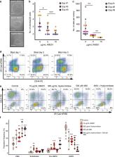
          The endothelial to haematopoietic transition (EHT) is the process whereby haemogenic endothelium differentiates into haematopoietic stem and progenitor cells (HSPCs). The intermediary steps of this process are unclear, in particular the identity of endothelial cells that give rise to HSPCs is unknown. Using single-cell transcriptome analysis and antibody screening, we identify CD44 as a marker of EHT enabling us to isolate robustly the different stages of EHT in the aorta-gonad-mesonephros (AGM) region. This allows us to provide a detailed phenotypical and transcriptional profile of CD44-positive arterial endothelial cells from which HSPCs emerge. They are characterized with high expression of genes related to Notch signalling, TGFbeta/BMP antagonists, a downregulation of genes related to glycolysis and the TCA cycle, and a lower rate of cell cycle. Moreover, we demonstrate that by inhibiting the interaction between CD44 and its ligand hyaluronan, we can block EHT, identifying an additional regulator of HSPC development.

          Abstract

          The endothelial to haematopoietic transition (EHT) is the process where haemogenic endothelium differentiates into haematopoietic stem and progenitor cells (HSPCs). Here the authors use single cell transcriptomics and antibody screening to identify CD44 as a marker of EHT that is required for EHT and HSPC development.

          Related collections

          Most cited references35

          • Record: found
          • Abstract: found
          • Article: not found

          Autophagy maintains the metabolism and function of young and old (hematopoietic) stem cells

          With age, hematopoietic stem cells (HSCs) lose their ability to regenerate the blood system, and promote disease development. Autophagy is associated with health and longevity, and is critical for protecting HSCs from metabolic stress. Here, we show that loss of autophagy in HSCs causes accumulation of mitochondria and an activated metabolic state, which drives accelerated myeloid differentiation mainly through epigenetic deregulations, and impairs HSC self-renewal activity and regenerative potential. Strikingly, the majority of HSCs in aged mice share these altered metabolic and functional features. However, ~ 1/3 of aged HSCs exhibit high autophagy levels and maintain a low metabolic state with robust long-term regeneration potential similar to healthy young HSCs. Our results demonstrate that autophagy actively suppresses HSC metabolism by clearing active, healthy mitochondria to maintain quiescence and stemness, and becomes increasingly necessary with age to preserve the regenerative capacity of old HSCs.
            Bookmark
            • Record: found
            • Abstract: found
            • Article: not found

            Hematopoietic stem cells derive directly from aortic endothelium during development

            A major goal of regenerative medicine is to instruct formation of multipotent, tissue-specific stem cells from induced pluripotent stem cells (iPSCs) for cell replacement therapies. Generation of hematopoietic stem cells (HSCs) from iPSCs or embryonic stem cells (ESCs) is not currently possible, however, necessitating a better understanding of how HSCs normally arise during embryonic development. We previously showed that hematopoiesis occurs through four distinct waves during zebrafish development, with HSCs arising in the final wave in close association with the dorsal aorta. Recent reports have suggested that murine HSCs derive from hemogenic endothelial cells (ECs) lining the aortic floor1,2. Additional in vitro studies have similarly suggested that the hematopoietic progeny of ESCs arise through intermediates with endothelial potential3,4. In this report, we have utilized the unique strengths of the zebrafish embryo to image directly the birth of HSCs from the ventral wall of the dorsal aorta. Utilizing combinations of fluorescent reporter transgenes, confocal timelapse microscopy and flow cytometry, we have identified and isolated the stepwise intermediates as aortic hemogenic endothelium transitions to nascent HSCs. Finally, using a permanent lineage tracing strategy, we demonstrate that the HSCs generated from hemogenic endothelium are the lineal founders of the adult hematopoietic system.
              Bookmark
              • Record: found
              • Abstract: found
              • Article: not found

              In vivo imaging of haematopoietic cells emerging from the mouse aortic endothelium.

              Haematopoietic stem cells (HSCs), responsible for blood production in the adult mouse, are first detected in the dorsal aorta starting at embryonic day 10.5 (E10.5). Immunohistological analysis of fixed embryo sections has revealed the presence of haematopoietic cell clusters attached to the aortic endothelium where HSCs might localize. The origin of HSCs has long been controversial and several candidates of the direct HSC precursors have been proposed (for review see ref. 7), including a specialized endothelial cell population with a haemogenic potential. Such cells have been described both in vitro in the embryonic stem cell (ESC) culture system and retrospectively in vivo by endothelial lineage tracing and conditional deletion experiments. Whether the transition from haemogenic endothelium to HSC actually occurs in the mouse embryonic aorta is still unclear and requires direct and real-time in vivo observation. To address this issue we used time-lapse confocal imaging and a new dissection procedure to visualize the deeply located aorta. Here we show the dynamic de novo emergence of phenotypically defined HSCs (Sca1(+), c-kit(+), CD41(+)) directly from ventral aortic haemogenic endothelial cells.
                Bookmark

                Author and article information

                Contributors
                christophe.lancrin@embl.it
                Journal
                Nat Commun
                Nat Commun
                Nature Communications
                Nature Publishing Group UK (London )
                2041-1723
                29 January 2020
                29 January 2020
                2020
                : 11
                : 586
                Affiliations
                [1 ]ISNI 0000 0004 0627 3632, GRID grid.418924.2, European Molecular Biology Laboratory, , EMBL Rome - Epigenetics and Neurobiology Unit, ; via E. Ramarini 32, 00015 Monterotondo, Italy
                [2 ]ISNI 0000 0004 0606 5382, GRID grid.10306.34, Wellcome Trust Sanger Institute, ; Wellcome Genome Campus, Hinxton, UK
                [3 ]ISNI 0000 0000 9709 7726, GRID grid.225360.0, European Molecular Biology Laboratory, , EMBL-EBI, ; Wellcome Genome Campus, Hinxton, Cambridgeshire CB10 1SD UK
                [4 ]ISNI 0000 0004 0495 846X, GRID grid.4709.a, European Molecular Biology Laboratory, , Structural and Computational Biology Unit, ; Meyerhofstrasse 1, 69117 Heidelberg, Germany
                [5 ]ISNI 0000000092721542, GRID grid.18763.3b, Moscow Institute of Physics and Technology, ; Institutskii Per. 9, Moscow Region, Dolgoprudny, 141700 Russia
                [6 ]European Molecular Biology Laboratory, Genomics Core Facility, Meyerhofstrasse 1, 69117 Heidelberg, Germany
                [7 ]ISNI 000000041936754X, GRID grid.38142.3c, Present Address: Stem Cell and Regenerative Biology Department, , Harvard University, ; 7 Divinity Avenue, Cambridge, MA 02138 USA
                [8 ]ISNI 0000000107068890, GRID grid.20861.3d, Present Address: Pachter Lab, Division of Biology and Biological Engineering, , California Institute of Technology, ; 1200 East California Boulevard, Pasadena, CA USA
                [9 ]ISNI 0000 0004 0491 4256, GRID grid.429509.3, Present Address: Max Planck Institute of Immunobiology and Epigenetics, ; Stübeweg 51, D-79108 Freiburg, Germany
                [10 ]Present Address: Institut für Medizinische Statistik und Bioinformatik, Bachemer Strasse 86, 50931 Köln, Germany
                [11 ]ISNI 0000 0001 0728 0170, GRID grid.10825.3e, Present Address: Department of Biochemistry and Molecular Biology, , The University of Southern Denmark, Danish Institute for Advanced Study, ; Campusvej 55, 5230 Odense M, Denmark
                Author information
                http://orcid.org/0000-0003-4013-6343
                http://orcid.org/0000-0002-9217-2330
                http://orcid.org/0000-0002-9264-1280
                http://orcid.org/0000-0002-0352-2547
                http://orcid.org/0000-0002-6166-8640
                http://orcid.org/0000-0002-6294-6366
                http://orcid.org/0000-0003-0028-7374
                Article
                14171
                10.1038/s41467-019-14171-5
                6989687
                31996681
                296b4325-b16b-412d-8ddc-966c531e790b
                © The Author(s) 2020

                Open Access This article is licensed under a Creative Commons Attribution 4.0 International License, which permits use, sharing, adaptation, distribution and reproduction in any medium or format, as long as you give appropriate credit to the original author(s) and the source, provide a link to the Creative Commons license, and indicate if changes were made. The images or other third party material in this article are included in the article’s Creative Commons license, unless indicated otherwise in a credit line to the material. If material is not included in the article’s Creative Commons license and your intended use is not permitted by statutory regulation or exceeds the permitted use, you will need to obtain permission directly from the copyright holder. To view a copy of this license, visit http://creativecommons.org/licenses/by/4.0/.

                History
                : 27 July 2018
                : 18 December 2019
                Categories
                Article
                Custom metadata
                © The Author(s) 2020

                Uncategorized
                computational biology and bioinformatics,haematopoietic stem cells,transcriptomics

                Comments

                Comment on this article