9
views
0
recommends
+1 Recommend
0 collections
    0
    shares
      • Record: found
      • Abstract: found
      • Article: found
      Is Open Access

      Inhibition of CTGF ameliorates peritoneal fibrosis through suppression of fibroblast and myofibroblast accumulation and angiogenesis

      research-article

      Read this article at

      Bookmark
          There is no author summary for this article yet. Authors can add summaries to their articles on ScienceOpen to make them more accessible to a non-specialist audience.

          Abstract

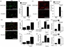
          Peritoneal fibrosis (PF) is a serious complication in various clinical settings, but the mechanisms driving it remain to be fully determined. Connective tissue growth factor (CTGF) is known to regulate fibroblast activities. We therefore examined if CTGF inhibition has anti-fibrotic effects in PF. PF was induced by repetitive intraperitoneal injections of chlorhexidine gluconate (CG) in mice with type I pro-collagen promoter-driven green fluorescent protein (GFP) expression to identify fibroblasts. FG-3019, an anti-CTGF monoclonal antibody, was used to inhibit CTGF. CG-induced PF was significantly attenuated in FG-3019-treated mice. CG challenges induced marked accumulations of proliferating fibroblasts and of myofibroblasts, which were both reduced by FG-3019. Levels of peritoneal CTGF expression were increased by CG challenges, and suppressed in FG-3019-treated mice. FG-3019 treatment also reduced the number of CD31 + vessels and VEGF-A-positive cells in fibrotic peritoneum. In vitro studies using NIH 3T3 fibroblasts and peritoneal mesothelial cells (PMCs) showed that CTGF blockade suppressed TGF-β 1-induced fibroblast proliferation and myofibroblast differentiation, PMC mesothelial-to-mesenchymal transition, and VEGF-A production. These findings suggest that the inhibition of CTGF by FG-3019 might be a novel treatment for PF through the regulation of fibroblast and myofibroblast accumulation and angiogenesis.

          Related collections

          Most cited references42

          • Record: found
          • Abstract: found
          • Article: not found

          Common and unique mechanisms regulate fibrosis in various fibroproliferative diseases.

          Fibroproliferative diseases, including the pulmonary fibroses, systemic sclerosis, liver cirrhosis, cardiovascular disease, progressive kidney disease, and macular degeneration, are a leading cause of morbidity and mortality and can affect all tissues and organ systems. Fibrotic tissue remodeling can also influence cancer metastasis and accelerate chronic graft rejection in transplant recipients. Nevertheless, despite its enormous impact on human health, there are currently no approved treatments that directly target the mechanism(s) of fibrosis. The primary goals of this Review series on fibrotic diseases are to discuss some of the major fibroproliferative diseases and to identify the common and unique mechanisms of fibrogenesis that might be exploited in the development of effective antifibrotic therapies.
            Bookmark
            • Record: found
            • Abstract: found
            • Article: not found

            The lysophosphatidic acid receptor LPA1 links pulmonary fibrosis to lung injury by mediating fibroblast recruitment and vascular leak.

            Aberrant wound-healing responses to injury have been implicated in the development of pulmonary fibrosis, but the mediators directing these pathologic responses have yet to be fully identified. We show that lysophosphatidic acid levels increase in bronchoalveolar lavage fluid following lung injury in the bleomycin model of pulmonary fibrosis, and that mice lacking one of its receptors, LPA1, are markedly protected from fibrosis and mortality in this model. The absence of LPA1 led to reduced fibroblast recruitment and vascular leak, two responses that may be excessive when injury leads to fibrosis rather than to repair, whereas leukocyte recruitment was preserved during the first week after injury. In persons with idiopathic pulmonary fibrosis, lysophosphatidic acid levels in bronchoalveolar lavage fluid were also increased, and inhibition of LPA1 markedly reduced fibroblast responses to the chemotactic activity of this fluid. LPA1 therefore represents a new therapeutic target for diseases in which aberrant responses to injury contribute to fibrosis, such as idiopathic pulmonary fibrosis.
              Bookmark
              • Record: found
              • Abstract: found
              • Article: not found

              Morphologic changes in the peritoneal membrane of patients with renal disease.

              This study examined the morphologic features of the parietal peritoneal membranes of 130 patients undergoing peritoneal dialysis (PD) and compared them with the features of the peritoneal membranes of normal individuals, uremic predialysis patients, and patients undergoing hemodialysis. The median thickness of the submesothelial compact collagenous zone was 50 microm for normal subjects, 140 microm for uremic patients, 150 microm for patients undergoing hemodialysis, and 270 microm for patients undergoing PD (P 97 mo, 700 microm (n = 19)]. Vascular changes included progressive subendothelial hyalinization, with luminal narrowing or obliteration. These changes were absent in samples from normal subjects but were present in 28% of samples from uremic patients and 56% of biopsies from patients undergoing PD. In the PD group, the prevalence of vasculopathy increased significantly with therapy duration (P = 0.0001). The density of blood vessels per unit length of peritoneum was significantly higher for patients with membrane failure and was correlated with the degree of fibrosis (P = 0.01). For the first time, a comprehensive cross-sectional analysis of the morphologic changes in the peritoneal membranes of patients undergoing PD is provided. The infrequency of fibrosis in the absence of vasculopathy suggests that vasculopathy may predispose patients to the development of fibrosis. This study provides a sufficiently large cohort of samples to allow structure-function relationships to be established, as well as providing a repository of tissue for further studies.
                Bookmark

                Author and article information

                Contributors
                norin0826@yahoo.co.jp
                Journal
                Sci Rep
                Sci Rep
                Scientific Reports
                Nature Publishing Group UK (London )
                2045-2322
                14 July 2017
                14 July 2017
                2017
                : 7
                : 5392
                Affiliations
                [1 ]ISNI 0000 0004 0615 9100, GRID grid.412002.5, Division of Nephrology, , Kanazawa University Hospital, ; Kanazawa, 920-8641 Japan
                [2 ]ISNI 0000 0004 0615 9100, GRID grid.412002.5, Division of Blood Purification, , Kanazawa University Hospital, ; Kanazawa, 920-8641 Japan
                [3 ]ISNI 0000 0001 2308 3329, GRID grid.9707.9, Department of Nephrology and Laboratory Medicine, Institute of Medical, Pharmaceutical and Health Sciences, , Kanazawa University, ; Kanazawa, 920-8641 Japan
                [4 ]ISNI 0000 0004 0409 3312, GRID grid.421404.7, , FibroGen, Inc., ; San Francisco, CA 94158 California USA
                [5 ]ISNI 0000 0001 2308 3329, GRID grid.9707.9, Department of System Biology, Institute of Medical, Pharmaceutical and Health Sciences, , Kanazawa University, ; Kanazawa, 920-8641 Japan
                [6 ]ISNI 000000041936754X, GRID grid.38142.3c, Center for Immunology and Inflammatory Diseases, , Harvard Medical School, ; Boston, MA 02114 USA
                [7 ]ISNI 000000041936754X, GRID grid.38142.3c, Division of Pulmonary and Critical Care Medicine, Massachusetts General Hospital, , Harvard Medical School, ; Boston, MA 02114 USA
                Author information
                http://orcid.org/0000-0001-8847-6051
                Article
                5624
                10.1038/s41598-017-05624-2
                5511333
                28710437
                2ba4c9cf-9f8f-464f-b2b3-97067d171d38
                © The Author(s) 2017

                Open Access This article is licensed under a Creative Commons Attribution 4.0 International License, which permits use, sharing, adaptation, distribution and reproduction in any medium or format, as long as you give appropriate credit to the original author(s) and the source, provide a link to the Creative Commons license, and indicate if changes were made. The images or other third party material in this article are included in the article’s Creative Commons license, unless indicated otherwise in a credit line to the material. If material is not included in the article’s Creative Commons license and your intended use is not permitted by statutory regulation or exceeds the permitted use, you will need to obtain permission directly from the copyright holder. To view a copy of this license, visit http://creativecommons.org/licenses/by/4.0/.

                History
                : 27 March 2017
                : 31 May 2017
                Categories
                Article
                Custom metadata
                © The Author(s) 2017

                Uncategorized
                Uncategorized

                Comments

                Comment on this article