50
views
0
recommends
+1 Recommend
0 collections
    0
    shares
      • Record: found
      • Abstract: found
      • Article: found
      Is Open Access

      Cellular correlates of longitudinal diffusion tensor imaging of axonal degeneration following hypoxic–ischemic cerebral infarction in neonatal rats

      research-article

      Read this article at

      Bookmark
          There is no author summary for this article yet. Authors can add summaries to their articles on ScienceOpen to make them more accessible to a non-specialist audience.

          Abstract

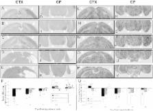
          Ischemically damaged brain can be accompanied by secondary degeneration of associated axonal connections e.g. Wallerian degeneration. Diffusion tensor imaging (DTI) is widely used to investigate axonal injury but the cellular correlates of many of the degenerative changes remain speculative. We investigated the relationship of DTI of directly damaged cerebral cortex and secondary axonal degeneration in the cerebral peduncle with cellular alterations in pan-axonal neurofilament staining, myelination, reactive astrocytes, activation of microglia/macrophages and neuronal cell death. DTI measures (axial, radial and mean diffusivity, and fractional anisotropy (FA)) were acquired at hyperacute (3 h), acute (1 and 2 d) and chronic (1 and 4 week) times after transient cerebral hypoxia with unilateral ischemia in neonatal rats. The tissue pathology underlying ischemic and degenerative responses had a complex relationship with DTI parameters. DTI changes at hyperacute and subacute times were smaller in magnitude and tended to be transient and/or delayed in cerebral peduncle compared to cerebral cortex. In cerebral peduncle by 1 d post-insult, there were reductions in neurofilament staining corresponding with decreases in parallel diffusivity which were more sensitive than mean diffusivity in detecting axonal changes. Ipsilesional reductions in FA within cerebral peduncle were robust in detecting both early and chronic degenerative responses. At one or four weeks post-insult, radial diffusivity was increased ipsilaterally in the cerebral peduncle corresponding to pathological evidence of a lack of ontogenic myelination in this region. The detailed differences in progression and magnitude of DTI and histological changes reported provide a reference for identifying the potential contribution of various cellular responses to FA, and, parallel, radial, and mean diffusivity.

          Highlights

          • Diffusion tensor imaging (DTI) widely used; cellular correlates often speculative

          • Studied longitudinal DTI and histological changes following hypoxia–ischemia

          • Compared neonatal cortex changes to those in degenerating cerebral peduncle

          • DTI and cellular changes were often transient or delayed in cerebral peduncle.

          • This provides a reference for potential cellular contributions to DTI changes.

          Related collections

          Most cited references30

          • Record: found
          • Abstract: found
          • Article: not found

          Fluoro-Jade B: a high affinity fluorescent marker for the localization of neuronal degeneration.

          Fluoro-Jade B, like its predecessor Fluoro-Jade, is an anionic fluorescein derivative useful for the histological staining of neurons undergoing degeneration. However, Fluoro-Jade B has an even greater specific affinity for degenerating neurons. This notion is supported by the conspicuous staining of degenerating neuronal elements with minimal background staining. This improved signal-to-noise ratio means that fine neuronal processes including distal dendrites, axons and axon terminals can be more readily detected and documented. Although the staining time and dye concentration are reduced, the method is as rapid, simple and reliable as the original Fluoro-Jade technique. Like Fluoro-Jade, Fluoro-Jade B is compatible with a number of other labeling procedures including immunofluorescent and fluorescent Nissl techniques.
            Bookmark
            • Record: found
            • Abstract: found
            • Article: not found

            A longitudinal diffusion tensor imaging study on Wallerian degeneration of corticospinal tract after motor pathway stroke.

            Wallerian degeneration of the corticospinal tract (CST) after motor pathway ischemic stroke can be characterized by diffusion tensor imaging (DTI). However, the dynamic evolution of the diffusion indices in the degenerated CST has not previously been completely identified. We investigated this dynamic evolution and the relationship between early changes of the diffusion indices in the degenerated CST and long-term clinical outcomes. DTI and neurological examinations were performed repeatedly in 9 patients with first-onset motor pathway subcortical infarction at 5 consecutive time points, i.e. within 1 week, at 2 weeks, 1 month, 3 months and 1 year. Using a region of interest method, we analyzed the ratios of the fractional anisotropy (rFA), mean diffusivity (rMD), primary eigenvalue (rlambda(1)) and transverse eigenvalue (rlambda(23)) between the affected and unaffected sides of the CSTs. We did not find any significant changes in the diffusion indices of the contralesional CSTs across time points. The rFA decreased monotonously during the first 3 months and then stabilized. The rMD increased after 2 weeks and stabilized after the third month. The rlambda(1) decreased during the first 2 weeks and then remained unchanged. The rlambda(23) increased during the first 3 months and then stabilized. We also found that the changes in the rFA between the first 2 time points were correlated with the NIHSS (P=0.00003) and the Motricity Indices (P=0.0004) after 1 year. Our results suggest that for patients with motor pathway stroke the diffusion indices in the degenerated CST stabilize within 3 months and that early changes in the rFA of the CST may predict long-term clinical outcomes.
              Bookmark
              • Record: found
              • Abstract: found
              • Article: not found

              Diffusion-tensor MR imaging and tractography: exploring brain microstructure and connectivity.

              Diffusion magnetic resonance (MR) imaging is evolving into a potent tool in the examination of the central nervous system. Although it is often used for the detection of acute ischemia, evaluation of directionality in a diffusion measurement can be useful in white matter, which demonstrates strong diffusion anisotropy. Techniques such as diffusion-tensor imaging offer a glimpse into brain microstructure at a scale that is not easily accessible with other modalities, in some cases improving the detection and characterization of white matter abnormalities. Diffusion MR tractography offers an overall view of brain anatomy, including the degree of connectivity between different regions of the brain. However, optimal utilization of the wide range of data provided with directional diffusion MR measurements requires careful attention to acquisition and postprocessing. This article will review the principles of diffusion contrast and anisotropy, as well as clinical applications in psychiatric, developmental, neurodegenerative, neoplastic, demyelinating, and other types of disease.
                Bookmark

                Author and article information

                Journal
                Neuroimage Clin
                Neuroimage Clin
                NeuroImage : Clinical
                Elsevier
                2213-1582
                7 August 2014
                7 August 2014
                2014
                : 6
                : 32-42
                Affiliations
                [a ]Hotchkiss Brain Institute, Faculty of Medicine, University of Calgary, Calgary, T2N 4N1, Canada
                [b ]Experimental Imaging Centre, Faculty of Medicine, University of Calgary, Calgary, AB T2N 4N1, Canada
                [c ]Departments of Physiology and Pharmacology, Faculty of Medicine, University of Calgary, Calgary, AB T2N 4N1, Canada
                [d ]Department of Clinical Neurosciences, Faculty of Medicine, University of Calgary, Calgary, AB T2N 4N1, Canada
                [e ]Department of Medical Physics and Informatics, School of Medicine, University of Szeged, Szeged, Hungary
                [f ]Department of Pediatrics, Alberta Children's Hospital Research Institute, Faculty of Medicine, University of Calgary, Calgary, AB T2N 4N1, Canada
                Author notes
                [* ]Corresponding author at: Depts of Physiology and Pharmacology, Clinical Neurosciences, and, Radiology, Univ. of Calgary, Teaching, Research and Wellness Building, Room P2E36, 3280 Hospital Drive N.W., Calgary, AB T2N 2T8, Canada.
                Article
                S2213-1582(14)00112-0
                10.1016/j.nicl.2014.08.003
                4215526
                25379414
                2be08605-d3f1-4b0a-80a7-4085e7bb1ea3
                © 2014 The Authors. Published by Elsevier Inc. All rights reserved.

                This is an open access article under the CC BY-NC-SA license (http://creativecommons.org/licenses/by-nc-sa/3.0/).

                History
                : 1 May 2014
                : 14 July 2014
                : 4 August 2014
                Categories
                Article

                magnetic resonance imaging,cerebral peduncle,neonatal cerebral hypoxia–ischemia,myelination,neurofilament,glial activation,mr, magnetic resonance,dti, diffusion tensor imaging,adc, apparent diffusion coefficient of water,fa, fractional anisotropy,gfap, glial fibrillary acidic protein,mbp, myelin basic protein

                Comments

                Comment on this article