2
views
0
recommends
+1 Recommend
0 collections
    0
    shares
      • Record: found
      • Abstract: found
      • Article: found
      Is Open Access

      Epithelial-Mesenchymal Transition Drives Three-Dimensional Morphogenesis in Mammalian Early Development

      research-article

      Read this article at

      Bookmark
          There is no author summary for this article yet. Authors can add summaries to their articles on ScienceOpen to make them more accessible to a non-specialist audience.

          Abstract

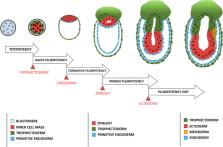
          From fertilization to onset of gastrulation, a mammalian embryo goes through several rounds of cellular morphogenesis resembling phenomena of epithelial-mesenchymal transition (EMT) and mesenchymal-epithelial transition (MET), collectively referred to as EMTs. How these EMT events play a role in shaping the three-dimensional (3-D) architecture of the developing embryo is not well-understood. In this review, we present a model in which cellular morphogenesis, represented primarily by dynamic changes in its epithelialization status, is the driving force of embryonic 3-D organization. This is achieved through the integration of three key components of mammalian early development, the pluripotency regulation, morphogenetic signaling, and biomechanical force anisotropy. Although cells in an early embryo do not exhibit full mesenchymal characteristics, our model underscores the importance of investigating molecular regulation of epithelial cell polarity and partial EMT/MET in understanding mammalian early development.

          Related collections

          Most cited references45

          • Record: found
          • Abstract: found
          • Article: not found

          Self-organization of the in vitro attached human embryo.

          Implantation of the blastocyst is a developmental milestone in mammalian embryonic development. At this time, a coordinated program of lineage diversification, cell-fate specification, and morphogenetic movements establishes the generation of extra-embryonic tissues and the embryo proper, and determines the conditions for successful pregnancy and gastrulation. Despite its basic and clinical importance, this process remains mysterious in humans. Here we report the use of a novel in vitro system to study the post-implantation development of the human embryo. We unveil the self-organizing abilities and autonomy of in vitro attached human embryos. We find human-specific molecular signatures of early cell lineage, timing, and architecture. Embryos display key landmarks of normal development, including epiblast expansion, lineage segregation, bi-laminar disc formation, amniotic and yolk sac cavitation, and trophoblast diversification. Our findings highlight the species-specificity of these developmental events and provide a new understanding of early human embryonic development beyond the blastocyst stage. In addition, our study establishes a new model system relevant to early human pregnancy loss. Finally, our work will also assist in the rational design of differentiation protocols of human embryonic stem cells to specific cell types for disease modelling and cell replacement therapy.
            Bookmark
            • Record: found
            • Abstract: found
            • Article: not found

            Membrane tension maintains cell polarity by confining signals to the leading edge during neutrophil migration.

            Little is known about how neutrophils and other cells establish a single zone of actin assembly during migration. A widespread assumption is that the leading edge prevents formation of additional fronts by generating long-range diffusible inhibitors or by sequestering essential polarity components. We use morphological perturbations, cell-severing experiments, and computational simulations to show that diffusion-based mechanisms are not sufficient for long-range inhibition by the pseudopod. Instead, plasma membrane tension could serve as a long-range inhibitor in neutrophils. We find that membrane tension doubles during leading-edge protrusion, and increasing tension is sufficient for long-range inhibition of actin assembly and Rac activation. Furthermore, reducing membrane tension causes uniform actin assembly. We suggest that tension, rather than diffusible molecules generated or sequestered at the leading edge, is the dominant source of long-range inhibition that constrains the spread of the existing front and prevents the formation of secondary fronts. Copyright © 2012 Elsevier Inc. All rights reserved.
              Bookmark
              • Record: found
              • Abstract: found
              • Article: not found

              A method to recapitulate early embryonic spatial patterning in human embryonic stem cells

              Embryos allocate cells to the three germ layers in a spatially ordered sequence. Human embryonic stem cells (hESCs) can generate the three germ layers in culture, however, differentiation is typically heterogeneous and spatially disordered. Here we show that geometric confinement is sufficient to trigger self-organized patterning in hESCs. In response to BMP4, these colonies reproducibly differentiate to an outer trophectoderm-like ring, an inner ectodermal circle and a ring of mesendoderm expressing primitive-streak markers in between. Fates are defined relative to the boundary with a fixed length scale: small colonies correspond to the outer layers of larger ones. Inhibitory signals limit the range of BMP4 signaling to the colony edge and induce a gradient of Activin/Nodal signaling that patterns mesendodermal fates. These results demonstrate that the intrinsic tendency of stem cells to make patterns can be harnessed by controlling colony geometries, and provide a quantitative assay for studying paracrine signaling.
                Bookmark

                Author and article information

                Contributors
                Journal
                Front Cell Dev Biol
                Front Cell Dev Biol
                Front. Cell Dev. Biol.
                Frontiers in Cell and Developmental Biology
                Frontiers Media S.A.
                2296-634X
                11 February 2021
                2021
                : 9
                : 639244
                Affiliations
                International Research Center for Medical Sciences (IRCMS), Kumamoto University , Kumamoto, Japan
                Author notes

                Edited by: Silvia Garagna, University of Pavia, Italy

                Reviewed by: Maria Cecilia Cirio, CONICET Institute of Physiology, Argentina; Isabelle Migeotte, Université libre de Bruxelles, Belgium; Alexandre Francou, Sloan Kettering Institute, United States

                *Correspondence: Guojun Sheng sheng@ 123456kumamoto-u.ac.jp

                This article was submitted to Cell Growth and Division, a section of the journal Frontiers in Cell and Developmental Biology

                †These authors have contributed equally to this work

                Article
                10.3389/fcell.2021.639244
                7905045
                2d1581a8-dbd3-4ff8-ac6a-76b630d3cebf
                Copyright © 2021 Ismagulov, Hamidi and Sheng.

                This is an open-access article distributed under the terms of the Creative Commons Attribution License (CC BY). The use, distribution or reproduction in other forums is permitted, provided the original author(s) and the copyright owner(s) are credited and that the original publication in this journal is cited, in accordance with accepted academic practice. No use, distribution or reproduction is permitted which does not comply with these terms.

                History
                : 08 December 2020
                : 06 January 2021
                Page count
                Figures: 5, Tables: 0, Equations: 0, References: 45, Pages: 7, Words: 4622
                Categories
                Cell and Developmental Biology
                Hypothesis and Theory

                emt,epiblast,pluripotency,polarity,gastrulation
                emt, epiblast, pluripotency, polarity, gastrulation

                Comments

                Comment on this article