5
views
0
recommends
+1 Recommend
0 collections
    0
    shares
      • Record: found
      • Abstract: found
      • Article: found
      Is Open Access

      LOXL1 modulates the malignant progression of colorectal cancer by inhibiting the transcriptional activity of YAP

      research-article

      Read this article at

      Bookmark
          There is no author summary for this article yet. Authors can add summaries to their articles on ScienceOpen to make them more accessible to a non-specialist audience.

          Abstract

          Background

          LOX-like 1 (LOXL1) is a lysyl oxidase, and emerging evidence has revealed its effect on malignant cancer progression. However, its role in colorectal cancer (CRC) and the underlying molecular mechanisms have not yet been elucidated.

          Methods

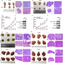
          LOXL1 expression in colorectal cancer was detected by immunohistochemistry, western blotting and real-time PCR. In vitro, colony formation, wound healing, migration and invasion assays were performed to investigate the effects of LOXL1 on cell proliferation, migration and invasion. In vivo, metastasis models and mouse xenografts were used to assess tumorigenicity and metastasis ability. Molecular biology experiments were utilized to reveal the underlying mechanisms by which LOXL1 modulates the Hippo pathway.

          Results

          LOXL1 was highly expressed in normal colon tissues compared with cancer tissues. In vitro , silencing LOXL1 in CRC cell lines dramatically enhanced migration, invasion, and colony formation, while overexpression of LOXL1 exerted the opposite effects. The results of the in vivo experiments demonstrated that the overexpression of LOXL1 in CRC cell lines drastically inhibited metastatic progression and tumour growth. Mechanistically, LOXL1 inhibited the transcriptional activity of Yes-associated protein (YAP) by interacting with MST1/2 and increasing the phosphorylation of MST1/2.

          Conclusions

          LOXL1 may function as an important tumour suppressor in regulating tumour growth, invasion and metastasis via negative regulation of YAP activity.

          Graphical abstract

          Related collections

          Most cited references29

          • Record: found
          • Abstract: found
          • Article: not found

          TEAD/TEF transcription factors utilize the activation domain of YAP65, a Src/Yes-associated protein localized in the cytoplasm.

          Mammals express four highly conserved TEAD/TEF transcription factors that bind the same DNA sequence, but serve different functions during development. TEAD-2/TEF-4 protein purified from mouse cells was associated predominantly with a novel TEAD-binding domain at the amino terminus of YAP65, a powerful transcriptional coactivator. YAP65 interacted specifically with the carboxyl terminus of all four TEAD proteins. Both this interaction and sequence-specific DNA binding by TEAD were required for transcriptional activation in mouse cells. Expression of YAP in lymphocytic cells that normally do not support TEAD-dependent transcription (e.g., MPC11) resulted in up to 300-fold induction of TEAD activity. Conversely, TEAD overexpression squelched YAP activity. Therefore, the carboxy-terminal acidic activation domain in YAP is the transcriptional activation domain for TEAD transcription factors. However, whereas TEAD was concentrated in the nucleus, excess YAP65 accumulated in the cytoplasm as a complex with the cytoplasmic localization protein, 14-3-3. Because TEAD-dependent transcription was limited by YAP65, and YAP65 also binds Src/Yes protein tyrosine kinases, we propose that YAP65 regulates TEAD-dependent transcription in response to mitogenic signals.
            Bookmark
            • Record: found
            • Abstract: found
            • Article: not found

            Mst1 and Mst2 protein kinases restrain intestinal stem cell proliferation and colonic tumorigenesis by inhibition of Yes-associated protein (Yap) overabundance.

            Ablation of the kinases Mst1 and Mst2, orthologs of the Drosophila antiproliferative kinase Hippo, from mouse intestinal epithelium caused marked expansion of an undifferentiated stem cell compartment and loss of secretory cells throughout the small and large intestine. Although median survival of mice lacking intestinal Mst1/Mst2 is 13 wk, adenomas of the distal colon are common by this age. Diminished phosphorylation, enhanced abundance, and nuclear localization of the transcriptional coactivator Yes-associated protein 1 (Yap1) is evident in Mst1/Mst2-deficient intestinal epithelium, as is strong activation of β-catenin and Notch signaling. Although biallelic deletion of Yap1 from intestinal epithelium has little effect on intestinal development, inactivation of a single Yap1 allele reduces Yap1 polypeptide abundance to nearly wild-type levels and, despite the continued Yap hypophosphorylation and preferential nuclear localization, normalizes epithelial structure. Thus, supraphysiologic Yap polypeptide levels are necessary to drive intestinal stem cell proliferation. Yap is overexpressed in 68 of 71 human colon cancers and in at least 30 of 36 colon cancer-derived cell lines. In colon-derived cell lines where Yap is overabundant, its depletion strongly reduces β-catenin and Notch signaling and inhibits proliferation and survival. These findings demonstrate that Mst1 and Mst2 actively suppress Yap1 abundance and action in normal intestinal epithelium, an antiproliferative function that frequently is overcome in colon cancer through Yap1 polypeptide overabundance. The dispensability of Yap1 in normal intestinal homeostasis and its potent proliferative and prosurvival actions when overexpressed in colon cancer make it an attractive therapeutic target.
              Bookmark
              • Record: found
              • Abstract: found
              • Article: not found

              Lysyl oxidase: an oxidative enzyme and effector of cell function.

              Lysyl oxidase (LOX) oxidizes the side chain of peptidyl lysine converting specific lysine residues to residues of alpha-aminoadipic-delta-semialdehyde. This posttranslational chemical change permits the covalent crosslinking of the component chains of collagen and those of elastin, thus stabilizing the fibrous deposits of these proteins in the extracellular matrix. Four LOX-like (LOXL) proteins with varying degrees of similarity to LOX have been described, constituting a family of related proteins. LOX is synthesized as a preproprotein which emerges from the cell as proLOX and then is processed to the active enzyme by proteolysis. In addition to elastin and collagen, LOX can oxidize lysine within a variety of cationic proteins, suggesting that its functions extend beyond its role in the stabilization of the extracellular matrix. Indeed, recent findings reveal that LOX and LOXL proteins markedly influence cell behavior including chemotactic responses, proliferation, and shifts between the normal and malignant phenotypes.
                Bookmark

                Author and article information

                Contributors
                hulin@zju.edu.cn
                wajing0123@163.com
                wylsoochow@qq.com
                Luckypeng5472@163.com
                wuchao123suda@163.com
                maobo98@qq.com
                emaruthip@gmail.com
                wangyuhong@suda.edu.cn
                chinyue@suda.edu.cn
                Journal
                Cell Commun Signal
                Cell Commun. Signal
                Cell Communication and Signaling : CCS
                BioMed Central (London )
                1478-811X
                10 September 2020
                10 September 2020
                2020
                : 18
                : 148
                Affiliations
                [1 ]GRID grid.263761.7, ISNI 0000 0001 0198 0694, Institutes of Biology and Medical Sciences, , Soochow University, ; Suzhou, China
                [2 ]GRID grid.429222.d, ISNI 0000 0004 1798 0228, Department of General surgery, , The First Affiliated Hospital of Soochow University, ; Suzhou, China
                [3 ]GRID grid.263761.7, ISNI 0000 0001 0198 0694, School of Biology and Basic Medical Science, , Soochow University, ; Suzhou, China
                [4 ]GRID grid.263488.3, ISNI 0000 0001 0472 9649, Department of Cell Biology and Genetics, Shenzhen key of Laboratory of Translational medicine of Tumor, , Shenzhen University Health science center, ; Shenzhen, China
                [5 ]GRID grid.429222.d, ISNI 0000 0004 1798 0228, Department of Pathology, , The First Affiliated Hospital of Soochow University, ; Suzhou, China
                Author information
                http://orcid.org/0000-0001-7864-0428
                Article
                639
                10.1186/s12964-020-00639-1
                7488294
                32912229
                2da46e15-af36-4610-aa5f-eb0268ea8491
                © The Author(s) 2020

                Open AccessThis article is licensed under a Creative Commons Attribution 4.0 International License, which permits use, sharing, adaptation, distribution and reproduction in any medium or format, as long as you give appropriate credit to the original author(s) and the source, provide a link to the Creative Commons licence, and indicate if changes were made. The images or other third party material in this article are included in the article's Creative Commons licence, unless indicated otherwise in a credit line to the material. If material is not included in the article's Creative Commons licence and your intended use is not permitted by statutory regulation or exceeds the permitted use, you will need to obtain permission directly from the copyright holder. To view a copy of this licence, visit http://creativecommons.org/licenses/by/4.0/. The Creative Commons Public Domain Dedication waiver ( http://creativecommons.org/publicdomain/zero/1.0/) applies to the data made available in this article, unless otherwise stated in a credit line to the data.

                History
                : 11 February 2020
                : 7 August 2020
                Funding
                Funded by: FundRef http://dx.doi.org/10.13039/501100001809, National Natural Science Foundation of China;
                Award ID: 81902969
                Award ID: 81902977
                Award Recipient :
                Funded by: FundRef http://dx.doi.org/10.13039/501100004608, Natural Science Foundation of Jiangsu Province;
                Award ID: BK20180839
                Award Recipient :
                Funded by: Natural Science Foundation of the Jiangsu Higher Education Institution of China
                Award ID: 18KJB320015
                Award Recipient :
                Funded by: FundRef http://dx.doi.org/10.13039/501100002858, China Postdoctoral Science Foundation;
                Award ID: 2018M630599, 2019T120454
                Award Recipient :
                Funded by: FundRef http://dx.doi.org/10.13039/501100012246, Priority Academic Program Development of Jiangsu Higher Education Institutions;
                Categories
                Research
                Custom metadata
                © The Author(s) 2020

                Cell biology
                colorectal cancer,loxl1,tumorigenesis,yes-associated protein
                Cell biology
                colorectal cancer, loxl1, tumorigenesis, yes-associated protein

                Comments

                Comment on this article