10
views
0
recommends
+1 Recommend
0 collections
    0
    shares
      • Record: found
      • Abstract: found
      • Article: found
      Is Open Access

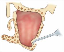
      Maxillary sinus augmentation

      review-article

      Read this article at

      Bookmark
          There is no author summary for this article yet. Authors can add summaries to their articles on ScienceOpen to make them more accessible to a non-specialist audience.

          Abstract

          The placement of endosseous implants in posterior edentulous maxilla is normally a challenging task in implant dentistry due to maxillary sinus pneumatization. Various sinus augmentation techniques have been used with impressive success rates aimed at developing these sites for implant placement. Knowledge of anatomy of maxillary sinus guides us not only in proper preoperative treatment planning but also helps us to avoid the possible complications that may arise during sinus augmentation procedure. This topic attracts a rising number of publications with most of them reporting results that suggest, the patients with atrophic maxillae requiring implant treatment can benefit considerably from the use of sinus augmentation. This article explains the basic techniques, namely, direct and indirect techniques used for maxillary sinus elevation and augmentation.

          Related collections

          Most cited references41

          • Record: found
          • Abstract: not found
          • Article: not found

          Grafting of the maxillary sinus floor with autogenous marrow and bone.

            Bookmark
            • Record: found
            • Abstract: found
            • Article: not found

            Maxillary and sinus implant reconstructions.

            H. Tatum (1986)
            The root and sinus series of the Omnii system have been used extensively since 1981. They are very versatile in their ability to be used within edentulous areas of the maxilla. Their design attempts to maximize the use of the available bone, and placement techniques allow the manipulation of bone to form sockets in otherwise deficient areas of bone. The root implants can be used as free-standing implants or as multiple abutments. The sinus implant is always used as an abutment. It may be used in conjunction with other implants or with natural abutments. Maxillary implants are not loaded until a 6-month healing time has elapsed following placement. An understanding of the different qualities of bone found in the maxilla is important to achieving the successful loading of these implants. Different times are required to allow physiologic loading in different qualities of maxillary bone. Restorative treatment is normally done with fixed bridge work, and the use of any type of stress breaker attachments is not recommended.
              Bookmark
              • Record: found
              • Abstract: found
              • Article: not found

              Anatomical aspects of sinus floor elevations.

              Inadequate bone height in the lateral part of the maxilla forms a contra-indication for implant surgery. This condition can be treated with an internal augmentation of the maxillary sinus floor. This sinus floor elevation, formerly called sinus lifting, consists of a surgical procedure in which a top hinge door in the lateral maxillary sinus wall is prepared and internally rotated to a horizontal position. The new elevated sinus floor, together with the inner maxillary mucosa, will create a space that can be filled with graft material. Sinus lift procedures depend greatly on fragile structures and anatomical variations. The variety of anatomical modalities in shape of the inner aspect of the maxillary sinus defines the surgical approach. Conditions such as sinus floor convolutions, sinus septum, transient mucosa swelling and narrow sinus may form a (usually relative) contra-indication for sinus floor elevation. Absolute contra-indications are maxillary sinus diseases (tumors) and destructive former sinus surgery (like the Caldwell-Luc operation). The lateral sinus wall is usually a thin bone plate, which is easily penetrated with rotating or sharp instruments. The fragile Schneiderian membrane plays an important role for the containment of the bonegraft. The surgical procedure of preparing the trap door and luxating it, together with the preparation of the sinus mucosa, may cause a mucosa tear. Usually, when these perforations are not too large, they will fold together when turning the trap door inward and upward, or they can be glued with a fibrin sealant, or they can be covered with a resorbable membrane. If the perforation is too large, a cortico-spongious block graft can be considered. However, in most cases the sinus floor elevation will be deleted. Perforations may also occur due to irregularities in the sinus floor or even due to immediate contact of sinus mucosa with oral mucosa. Obstruction of the antro-nasal foramen is, due to its high location, not a likely complication, nor is the occurrence of severe haemorrhages since the trap door is in the periphery of the supplying vessels. Apart from these two aspects, a number of anatomical considerations are described in connection with sinus floor elevation.
                Bookmark

                Author and article information

                Journal
                J Indian Soc Periodontol
                J Indian Soc Periodontol
                JISP
                Journal of Indian Society of Periodontology
                Medknow Publications & Media Pvt Ltd (India )
                0972-124X
                0975-1580
                Nov-Dec 2018
                : 22
                : 6
                : 468-473
                Affiliations
                [1] Department of Periodontology and Oral Implantology, M M College of Dental Sciences and Research, Ambala, Haryana, India
                [1 ] Department of Oral and Maxillofacial Surgery Implantology, M M College of Dental Sciences and Research, Ambala, Haryana, India
                [2 ] Private Practitioner, Mumbai, Maharashtra, India
                Author notes
                Address for correspondence: Dr. Shalu Chandna Bathla, H. No 782, Sector 13, Urban Estate, Karnal, Haryana, India. E-mail: periodonticsrevisited@ 123456rediffmail.com
                Article
                JISP-22-468
                10.4103/jisp.jisp_236_18
                6305100
                30631223
                2e13f839-3d52-4ad2-a309-f1dcc394b80f
                Copyright: © 2018 Indian Society of Periodontology

                This is an open access journal, and articles are distributed under the terms of the Creative Commons Attribution-NonCommercial-ShareAlike 4.0 License, which allows others to remix, tweak, and build upon the work non-commercially, as long as appropriate credit is given and the new creations are licensed under the identical terms.

                History
                : 09 April 2018
                : 01 June 2018
                Categories
                Review Article

                Dentistry
                crestal approach,direct lateral window,maxillary sinus,maxillary sinus augmentation,osteotome

                Comments

                Comment on this article