105
views
0
recommends
+1 Recommend
0 collections
    0
    shares
      • Record: found
      • Abstract: found
      • Article: not found

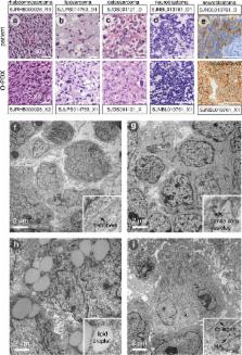
      Orthotopic Patient-Derived Xenografts of Pediatric Solid Tumors

      research-article

      Read this article at

      ScienceOpenPublisherPMC
      Bookmark
          There is no author summary for this article yet. Authors can add summaries to their articles on ScienceOpen to make them more accessible to a non-specialist audience.

          Abstract

          Pediatric solid tumors arise from endodermal, ectodermal, or mesodermal lineages 1 . Although the overall survival of children with solid tumors is 75%, that of children with recurrent disease is below 30% 2 . To capture the complexity and diversity of pediatric solid tumors and establish new models of recurrent disease, we developed a protocol to produce orthotopic patient-derived xenografts (O-PDXs) at diagnosis, recurrence, and autopsy. Tumor specimens were received from 168 patients, and 67 O-PDXs were established for 12 types of cancer. The origins of the O-PDX tumors were reflected in their gene-expression profiles and epigenomes. Genomic profiling of the tumors, including detailed clonal analysis, was performed to determine whether the clonal population in the xenograft recapitulated the patient’s tumor. We identified several drug vulnerabilities and showed that the combination of a WEE1 inhibitor (AZD1775), irinotecan, and vincristine can lead to complete response in multiple rhabdomyosarcoma O-PDX tumors in vivo.

          Related collections

          Most cited references15

          • Record: found
          • Abstract: not found
          • Article: not found

          Controlling the False Discovery Rate: A Practical and Powerful Approach to Multiple Testing

            Bookmark
            • Record: found
            • Abstract: found
            • Article: not found

            Organoid cultures derived from patients with advanced prostate cancer.

            The lack of in vitro prostate cancer models that recapitulate the diversity of human prostate cancer has hampered progress in understanding disease pathogenesis and therapy response. Using a 3D organoid system, we report success in long-term culture of prostate cancer from biopsy specimens and circulating tumor cells. The first seven fully characterized organoid lines recapitulate the molecular diversity of prostate cancer subtypes, including TMPRSS2-ERG fusion, SPOP mutation, SPINK1 overexpression, and CHD1 loss. Whole-exome sequencing shows a low mutational burden, consistent with genomics studies, but with mutations in FOXA1 and PIK3R1, as well as in DNA repair and chromatin modifier pathways that have been reported in advanced disease. Loss of p53 and RB tumor suppressor pathway function are the most common feature shared across the organoid lines. The methodology described here should enable the generation of a large repertoire of patient-derived prostate cancer lines amenable to genetic and pharmacologic studies. Copyright © 2014 Elsevier Inc. All rights reserved.
              Bookmark
              • Record: found
              • Abstract: not found
              • Article: not found

              Bioassay Analysis usingR

                Bookmark

                Author and article information

                Journal
                0410462
                6011
                Nature
                Nature
                Nature
                0028-0836
                1476-4687
                31 July 2017
                30 August 2017
                07 September 2017
                28 February 2018
                : 549
                : 7670
                : 96-100
                Affiliations
                [1 ]Department of Oncology, Memphis, Tennessee, 38105, USA
                [2 ]Department of Developmental Neurobiology, Memphis, Tennessee, 38105, USA
                [3 ]Department of Computational Biology, Memphis, Tennessee, 38105, USA
                [4 ]Department of Chemical Biology and Therapeutics, Memphis, Tennessee, 38105, USA
                [5 ]Department of Small Animal Imaging, Memphis, Tennessee, 38105, USA
                [6 ]Department of Pathology, Memphis, Tennessee, 38105, USA
                [7 ]Department of Preclinical Pharmacokinetics Shared Resource, Memphis, Tennessee, 38105, USA
                [8 ]Department of Biostatistics St. Jude Children’s Research Hospital, Memphis, Tennessee, 38105, USA
                [9 ]The Genome Institute, Washington University School of Medicine in St. Louis, St. Louis, Missouri 63108, USA
                [10 ]Department of Genetics, Washington University School of Medicine in St. Louis, St. Louis, Missouri 63108, USA
                [11 ]Siteman Cancer Center, Washington University School of Medicine in St. Louis, St. Louis, Missouri 63108, USA
                [12 ]Department of Medicine, Washington University School of Medicine in St. Louis, St. Louis, Missouri 63108, USA
                [13 ]Howard Hughes Medical Institute, Chevy Chase, Maryland 20815, USA
                [14 ]Department of Ophthalmology, University of Tennessee Health Science Center, Memphis, Tennessee, 38105, USA
                Author notes
                Correspondence and requests for materials should be addressed to: Michael A. Dyer, Department of Developmental Neurobiology, MS 323, St. Jude Children’s Research Hospital, 262 Danny Thomas Place, Memphis, TN, 38105-3678, USA, Phone: (901) 595-2257; Fax: (901) 595-3143; michael.dyer@ 123456stjude.org , http://stjude.org/CSTN
                [*]

                These authors contributed equally.

                Article
                NIHMS893713
                10.1038/nature23647
                5659286
                28854174
                2f9ec88d-066c-4db6-97e6-56b0ed5edee0

                Users may view, print, copy, and download text and data-mine the content in such documents, for the purposes of academic research, subject always to the full Conditions of use: http://www.nature.com/authors/editorial_policies/license.html#terms

                Reprints and permissions information is available at www.nature.com/reprints.

                History
                Categories
                Article

                Uncategorized
                Uncategorized

                Comments

                Comment on this article