33
views
0
recommends
+1 Recommend
0 collections
    0
    shares
      • Record: found
      • Abstract: found
      • Article: found
      Is Open Access

      Effects of chronic exposure to sodium arsenite on hypothalamo-pituitary-testicular activities in adult rats: possible an estrogenic mode of action

      research-article
      1 , 2 , , 2 , 1
      Reproductive Biology and Endocrinology
      BioMed Central

      Read this article at

      Bookmark
          There is no author summary for this article yet. Authors can add summaries to their articles on ScienceOpen to make them more accessible to a non-specialist audience.

          Abstract

          Background

          Inorganic arsenic is a major water pollutant and a known human carcinogen that has a suppressive influence on spermatogenesis and androgenesis in male reproductive system. However, the actual molecular events resulting in male reproductive dysfunctions from exposure to arsenic remain unclear. In this context, we evaluated the mode of action of chronic oral exposure of sodium arsenite on hypothalamo-pituitary- testicular activities in mature male albino rats.

          Methods

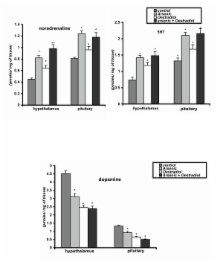
          The effect of chronic oral exposure to sodium arsenite (5 mg/kg body weight/day) via drinking water without or with hCG (5 I.U./kg body weight/day) and oestradiol (25 micrograms oestradiol 3-benzoate suspended in 0.25 ml olive oil/rat/day) co-treatments for 6 days a week for 4 weeks (about the duration of two spermatogenic cycle) was evaluated in adult male rats. Changes in paired testicular weights, quantitative study of different varieties of germ cells at stage VII of spermatogenic cycle, epididymal sperm count, circulatory concentrations of hormones (LH, FSH, testosterone and corticosterone), testicular activities of delta 5, 3beta-hydroxysteroid dehydrogenase (delta 5, 3beta-HSD), 17 beta-hydroxysteroid dehydrogenase (17 beta-HSD), sorbitol dehydrogenase (SDH), acid phosphatase (ACP), alkaline phosphatase (ALP), and lactate dehydrogenase (LDH), as well as the levels of biogenic amines (dopamine, noradrenaline and 5-hydroxytryptamine (5-HT)) in the hypothalamus and pituitary were monitored in this study. Hormones were assayed by radioimmuno- assay or enzyme- linked immunosorbent assay and the enzymes were estimated after spectrophotometry as well as the biogenic amines by HPLC electrochemistry.

          Results

          Sodium arsenite treatment resulted in: decreased paired testicular weights; epididymal sperm count; plasma LH, FSH, testosterone and testicular testosterone concentrations; and increased plasma concentration of corticosterone. Testicular enzymes such as delta 5, 3 beta-HSD, 17 beta-HSD, and sorbitol dehydrogenase (SDH) were significantly decreased, but those of acid phosphatase (ACP), alkaline phosphatase (ALP), and lactate dehydrogenase (LDH) were significantly increased. A decrease in dopamine or an increase in noradrenaline and 5-HT in hypothalamus and pituitary were also noted after arsenic exposure. Histological evaluation revealed extensive degeneration of different varieties of germ cells at stage VII of spermatogenic cycle in arsenic exposed rats. Administration of human chorionic gonadotrophin (hCG) along with sodium arsenite partially prevented the degeneration of germ cells and enhanced paired testicular weights, epididymal sperm count, plasma and intratesticular testosterone concentrations, activities of delta 5, 3beta-HSD, 17 beta-HSD and sorbitol dehydrogenase along with diminution in the activities of ACP, ALP and LDH. Since many of the observed arsenic effects could be enhanced by oestradiol, it is suggested that arsenic might somehow acts through an estrogenic mode of action.

          Conclusion

          The results indicate that arsenic causes testicular toxicity by germ cell degeneration and inhibits androgen production in adult male rats probably by affecting pituitary gonadotrophins. Estradiol treatment has been associated with similar effects on pituitary testicular axis supporting the hypothesis that arsenite might somehow act through an estrogenic mode of action.

          Related collections

          Most cited references86

          • Record: found
          • Abstract: found
          • Article: not found

          Estrogen and spermatogenesis.

          Although it has been known for many years that estrogen administration has deleterious effects on male fertility, data from transgenic mice deficient in estrogen receptors or aromatase point to an essential physiological role for estrogen in male fertility. This review summarizes the current knowledge on the localization of estrogen receptors and aromatase in the testis in an effort to understand the likely sites of estrogen action. The review also discusses the many studies that have used models employing the administration of estrogenic substances to show that male fertility is responsive to estrogen, thus providing a mechanism by which inappropriate exposure to estrogenic substances may cause adverse effects on spermatogenesis and male fertility. The reproductive phenotypes of mice deficient in estrogen receptors alpha and/or beta and aromatase are also compared to evaluate the physiological role of estrogen in male fertility. The review focuses on the effects of estrogen administration or deprivation, primarily in rodents, on the hypothalamo-pituitary-testis axis, testicular function (including Leydig cell, Sertoli cell, and germ cell development and function), and in the development and function of the efferent ductules and epididymis. The requirement for estrogen in normal male sexual behavior is also reviewed, along with the somewhat limited data on the fertility of men who lack either the capacity to produce or respond to estrogen. This review highlights the ability of exogenous estrogen exposure to perturb spermatogenesis and male fertility, as well as the emerging physiological role of estrogens in male fertility, suggesting that, in this local context, estrogenic substances should also be considered "male hormones."
            Bookmark
            • Record: found
            • Abstract: found
            • Article: not found

            Transplacental carcinogenicity of inorganic arsenic in the drinking water: induction of hepatic, ovarian, pulmonary, and adrenal tumors in mice.

            Arsenic is a known human carcinogen, but development of rodent models of inorganic arsenic carcinogenesis has been problematic. Since gestation is often a period of high sensitivity to chemical carcinogenesis, we performed a transplacental carcinogenicity study in mice using inorganic arsenic. Groups (n = 10) of pregnant C3H mice were given drinking water containing sodium arsenite (NaAsO(2)) at 0 (control), 42.5, and 85 ppm arsenite ad libitum from day 8 to 18 of gestation. These doses were well tolerated and body weights of the dams during gestation and of the offspring subsequent to birth were not reduced. Dams were allowed to give birth, and offspring were weaned at 4 weeks and then put into separate gender-based groups (n = 25) according to maternal exposure level. The offspring received no additional arsenic treatment. The study lasted 74 weeks in males and 90 weeks in females. A complete necropsy was performed on all mice and tissues were examined by light microscopy in a blind fashion. In male offspring, there was a marked increase in hepatocellular carcinoma incidence in a dose- related fashion (control, 12%; 42.5 ppm, 38%; 85 ppm, 61%) and in liver tumor multiplicity (tumors per liver; 5.6-fold over control at 85 ppm). In males, there was also a dose-related increase in adrenal tumor incidence and multiplicity. In female offspring, dose-related increases occurred in ovarian tumor incidence (control, 8%; 42.5 ppm, 26%; 85 ppm, 38%) and lung carcinoma incidence (control, 0%; 42.5 ppm, 4%; 85 ppm, 21%). Arsenic exposure also increased the incidence of proliferative lesions of the uterus and oviduct. These results demonstrate that oral inorganic arsenic exposure, as a single agent, can induce tumor formation in rodents and establishes inorganic arsenic as a complete transplacental carcinogen in mice. The development of this rodent model of inorganic arsenic carcinogenesis has important implications in defining the mechanism of action for this common environmental carcinogen. Copyright 2003 Elsevier Science (USA)
              Bookmark
              • Record: found
              • Abstract: found
              • Article: not found

              Induction of oxyradicals by arsenic: implication for mechanism of genotoxicity.

              Although arsenic is a well-established human carcinogen, the mechanisms by which it induces cancer remain poorly understood. We previously showed arsenite to be a potent mutagen in human-hamster hybrid (A(L)) cells, and that it induces predominantly multilocus deletions. We show here by confocal scanning microscopy with the fluorescent probe 5',6'-chloromethyl-2',7'-dichlorodihydrofluorescein diacetate that arsenite induces, within 5 min after treatment, a dose-dependent increase of up to 3-fold in intracellular oxyradical production. Concurrent treatment of cells with arsenite and the radical scavenger DMSO reduced the fluorescent intensity to control levels. ESR spectroscopy with 4-hydroxy-2,2,6,6-tetramethyl-1-hydroxypiperidine (TEMPOL-H) as a probe in conjunction with superoxide dismutase and catalase to quench superoxide anions and hydrogen peroxide, respectively, indicates that arsenite increases the levels of superoxide-driven hydroxyl radicals in these cells. Furthermore, reducing the intracellular levels of nonprotein sulfhydryls (mainly glutathione) in A(L) cells with buthionine S-R-sulfoximine increases the mutagenic potential of arsenite by more than 5-fold. The data are consistent with our previous results with the radical scavenger DMSO, which reduced the mutagenicity of arsenic in these cells, and provide convincing evidence that reactive oxygen species, particularly hydroxyl radicals, play an important causal role in the genotoxicity of arsenical compounds in mammalian cells.
                Bookmark

                Author and article information

                Journal
                Reprod Biol Endocrinol
                Reproductive Biology and Endocrinology
                BioMed Central (London )
                1477-7827
                2006
                16 February 2006
                : 4
                : 9
                Affiliations
                [1 ]Department of veterinary Surgery and radiology, west Bengal University of Animal and Fishery Sciences, 37 and 68, K. B. Sarani, Calcutta- 700 037, West Bengal, India
                [2 ]Institute of Molecular Medicine, Bengal Intelligent Park Ltd., Sector-V, Salt Lake Electronics Complex, Calcutta-700 091, India
                Article
                1477-7827-4-9
                10.1186/1477-7827-4-9
                1397838
                16483355
                31a6e4cd-2f94-4007-9a9e-f98176e71963
                Copyright © 2006 Jana et al; licensee BioMed Central Ltd.

                This is an Open Access article distributed under the terms of the Creative Commons Attribution License ( http://creativecommons.org/licenses/by/2.0), which permits unrestricted use, distribution, and reproduction in any medium, provided the original work is properly cited.

                History
                : 17 November 2005
                : 16 February 2006
                Categories
                Research

                Human biology
                Human biology

                Comments

                Comment on this article