8
views
0
recommends
+1 Recommend
0 collections
    0
    shares
      • Record: found
      • Abstract: found
      • Article: found
      Is Open Access

      Peripheral Dopamine in Restless Legs Syndrome

      research-article

      Read this article at

      Bookmark
          There is no author summary for this article yet. Authors can add summaries to their articles on ScienceOpen to make them more accessible to a non-specialist audience.

          Abstract

          Objective/Background

          Restless Legs Syndrome (RLS) is a dopamine-dependent disorder characterized by a strong urge to move. The objective of this study was to evalulate blood levels of dopamine and other catecholamines and blood D2-subtype dopamine receptors (D2Rs) in RLS.

          Patients/Methods

          Dopamine levels in blood samples from age-matched unmedicated RLS subjects, medicated RLS subjects and Controls were evaluated with high performance liquid chromatography and dopamine D2R white blood cell (WBC) expression levels were determined with fluorescence-activated cell sorting and immunocytochemistry.

          Results

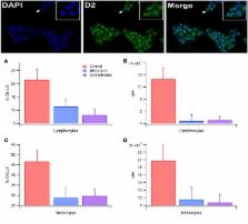
          Blood plasma dopamine levels, but not norepinepherine or epinephrine levels, were significantly increased in medicated RLS subjects vs unmedicated RLS subjects and Controls. The percentage of lymphocytes and monocytes expressing D2Rs differed between Control, RLS medicated and RLS unmedicated subjects. Total D2R expression in lymphocytes, but not monocytes, differed between Control, RLS medicated and RLS unmedicated subjects. D2Rs in lymphocytes, but not monocytes, were sensitive to dopamine in Controls only.

          Conclusion

          Downregulation of WBCs D2Rs occurs in RLS. This downregulation is not reversed by medication, although commonly used RLS medications increase plasma dopamine levels. The insensitivity of monocytes to dopamine levels, but their downregulation in RLS, may reflect their utility as a biomarker for RLS and perhaps brain dopamine homeostasis.

          Related collections

          Most cited references35

          • Record: found
          • Abstract: found
          • Article: not found

          Profound decreases in dopamine release in striatum in detoxified alcoholics: possible orbitofrontal involvement.

          The value of rewards (natural rewards and drugs) is associated with dopamine increases in the nucleus accumbens and varies as a function of context. The prefrontal cortex has been implicated in the context dependency of rewards and in the fixated high value that drugs have in addiction, although the mechanisms are not properly understood. Here we test the hypothesis that the prefrontal cortex regulates the value of rewards by modulating dopamine increases in nucleus accumbens and that this regulation is disrupted in addicted subjects. We used positron emission tomography to evaluate the activity of the prefrontal cortex (measuring brain glucose metabolism with [18F]fluorodeoxyglucose) and dopamine increases (measured with [11C]raclopride, a D2/D3 receptor ligand with binding that is sensitive to endogenous dopamine) induced by the stimulant drug methylphenidate in 20 controls and 20 detoxified alcoholics, most of whom smoked. In all subjects, methylphenidate significantly increased dopamine in striatum. In ventral striatum (where the nucleus accumbens is located) and in putamen, dopamine increases were associated with the rewarding effects of methylphenidate (drug liking and high) and were profoundly attenuated in alcoholics (70 and 50% lower than controls, respectively). In controls, but not in alcoholics, metabolism in orbitofrontal cortex (region involved with salience attribution) was negatively associated with methylphenidate-induced dopamine increases in ventral striatum. These results are consistent with the hypothesis that the orbitofrontal cortex modulates the value of rewards by regulating the magnitude of dopamine increases in the ventral striatum and that disruption of this regulation may underlie the decreased sensitivity to rewards in addicted subjects.
            Bookmark
            • Record: found
            • Abstract: found
            • Article: not found

            Human CD4+CD25+ regulatory T cells selectively express tyrosine hydroxylase and contain endogenous catecholamines subserving an autocrine/paracrine inhibitory functional loop.

            CD4+CD25+ regulatory T lymphocytes (Tregs) are specialized T cells playing a key role in the control of immune homeostasis. Here, we show that human Tregs constitutively express tyrosine hydroxylase (TH, EC 1.14.16.2), the rate-limiting enzyme in the synthesis of catecholamines, and contain substantial amounts of dopamine, norepinephrine, and epinephrine, which are released upon treatment with reserpine. Catecholamine release results in reduced production of interleukin-10 and transforming growth factor-beta by Tregs, and in down-regulation of Treg-dependent inhibition of effector T-lymphocyte (Teff) proliferation, which occurs without affecting the production of tumor necrosis factor-alpha or interferon-gamma. Tregs and Teffs express on the cell membrane both D1-like and D2-like dopaminergic receptors to a similar extent (12%-29% of the cells). Catecholamine-dependent down-regulation of Tregs is, however, selectively reversed by pharmacological blockade of dopaminergic D1-like receptors, which in Tregs only (and not in Teffs) are also expressed at the level of mRNA and are functionally coupled to intracellular production of cAMP. These findings indicate that in human Tregs endogenous catecholamines subserve an autocrine/paracrine loop involving dopaminergic pathways and resulting in down-regulation of Treg function.
              Bookmark
              • Record: found
              • Abstract: found
              • Article: not found

              Restless legs syndrome: revisiting the dopamine hypothesis from the spinal cord perspective.

              Restless legs syndrome (RLS) involves abnormal limb sensations that diminish with motor activity, worsen at rest, have a circadian peak in expression in the evening and at night, and can severely disrupt sleep. Primary treatment is directed at CNS dopaminergic systems, particularly activation of D(2)-like (D(2), D(3), and D(4)) receptors. Although RLS affects 2% to 15% of the general population, the neural circuitry contributing to RLS remains speculative, and there is currently no accepted animal model to enable detailed mechanistic analyses. Traditional views suggest that RLS arises from supraspinal sources which favor facilitation of the flexor reflex and emergence of the RLS phenotype. The authors forward the hypothesis that RLS reflects a dysfunction of the little-studied dorsoposterior hypothalamic dopaminergic A11 cell group. They assert that, as the sole source of spinal dopamine, reduced drive in this system can lead to spinal network changes wholly consistent with RLS. The authors summarize their recent investigations on spinal cord dopamine dysfunction that rely on lesions centered on A11, and on studies in D(3) receptor knockout (D(3)KO) mice. Excessive locomotor behavior is evident in both sets of animals, and D(3)KO mice exhibit facilitation rather than the expected depression of spinal reflexes in the presence of dopamine as well as a reversal in their circadian expression of the rate-limiting enzyme for dopamine synthesis, tyrosine hydroxylase. Taken together, these findings are consistent with an involvement of spinal dopamine dysfunction in the etiology of RLS, and they argue that the D(3)KO mouse might serve as a relevant animal model to study the underlying mechanisms of RLS.
                Bookmark

                Author and article information

                Contributors
                URI : http://frontiersin.org/people/u/463686
                URI : http://frontiersin.org/people/u/504238
                URI : http://frontiersin.org/people/u/515619
                URI : http://frontiersin.org/people/u/485531
                Journal
                Front Neurol
                Front Neurol
                Front. Neurol.
                Frontiers in Neurology
                Frontiers Media S.A.
                1664-2295
                15 March 2018
                2018
                : 9
                : 155
                Affiliations
                [1] 1Department of Exercise Sciences, Brigham Young University , Provo, UT, United States
                [2] 2Department of Psychology, Brigham Young University , Provo, UT, United States
                [3] 3Department of Microbiology and Molecular Biology, Brigham Young University , Provo, UT, United States
                Author notes

                Edited by: Huifang Shang, Sichuan University, China

                Reviewed by: Pedro Ribeiro, Universidade Federal do Rio de Janeiro, Brazil; Matteo Bologna, Sapienza Università di Roma, Italy

                *Correspondence: Ulrike H. Mitchell, rike_mitchell@ 123456byu.edu

                Specialty section: This article was submitted to Movement Disorders, a section of the journal Frontiers in Neurology

                Article
                10.3389/fneur.2018.00155
                5862810
                29599746
                31ffbc68-a6db-462a-ac89-c6c47f324aa2
                Copyright © 2018 Mitchell, Obray, Hunsaker, Garcia, Clarke, Hope and Steffensen.

                This is an open-access article distributed under the terms of the Creative Commons Attribution License (CC BY). The use, distribution or reproduction in other forums is permitted, provided the original author(s) and the copyright owner are credited and that the original publication in this journal is cited, in accordance with accepted academic practice. No use, distribution or reproduction is permitted which does not comply with these terms.

                History
                : 04 December 2017
                : 02 March 2018
                Page count
                Figures: 3, Tables: 1, Equations: 0, References: 40, Pages: 7, Words: 5433
                Categories
                Neuroscience
                Original Research

                Neurology
                willis–ekbom disease,da,d2 receptors,white blood cells,lymphocytes,monocytes
                Neurology
                willis–ekbom disease, da, d2 receptors, white blood cells, lymphocytes, monocytes

                Comments

                Comment on this article