- Record: found
- Abstract: found
- Article: found
Giant cell myocarditis in the CMR era
research-article
Tora Leong
1
,
,
Joyce Wong
1 ,
Alexandra Rice
1 ,
Mamdouh Zidan
1 ,
Andrew Hamilton
1 ,
Ben Ariff
1 ,
Ruth Chester
1 ,
Shelley L Rahman Haley
1 ,
Andrew Kelion
1 ,
Margaret M Burke
1 ,
Andrew G Mitchell
1 ,
Nicholas Banner
1 ,
Tarun K Mittal
1
1 February 2012
15th Annual SCMR Scientific Sessions
2-5 February 2012
Read this article at
There is no author summary for this article yet. Authors can add summaries to their articles on ScienceOpen to make them more accessible to a non-specialist audience.
Abstract
Summary
Our series of 5 cases of histologically-proven Giant cell myocarditis with concurrent
CMR shows a pattern of late gadolinium enhancement which tends to be widespread involving
all layers of the myocardium.
Background
Giant cell myocarditis (GCM) is a rare condition with paucity of data, particularly
on diagnosis, prognosis and morphological correlation. We sought to review cases of
histologically-proven GCM to examine their presentation and investigations, and in
particular, the potential role of cardiac magnetic resonance (CMR) imaging in the
diagnosis.
Methods
Cases of histologically-proven GCM presenting to our institution, a national transplant
center, who had concurrent CMR imaging were identified. CMR findings were evaluated
from the initial and follow-up scans for ventricular volumes, function, and late gadolinium
enhancement (LGE) on an advanced post-processing workstation.
Results
5 patients with histologically-proven GCM had CMR performed with 2 having repeat CMR.
4 out of the 5 patients were in cardiogenic shock at the time of biopsy (Patients
No: 1, 2, 4 and 5). Most patients were initially too unwell for CMR. For the 5 patients,
their times from presentation at our institution to time of endomyocardial biopsy
(and initiation of treatment soon after) were 0, 3, 11, 0 and 8 days respectively
while their times from presentation to first CMR were 12, 31, 9, 56 and 545 days respectively.
Table 1 summarizes the CMR findings of these 5 patients and their follow-up. They
had moderate to severe reduction in left ventricular (LV) systolic function largely
due to increase in end-systolic volumes (ESV) with the end-diastolic dimensions remaining
within normal limits. All patients had LGE affecting the myocardium of the LV with
multi-focal involvement of all layers of myocardium with no segmental predisposition.
3 patients also had LGE in the right ventricular (RV) myocardium. In the 2 patients
who had follow-up CMR, there was deterioration in LV EF due to increasing LV ESV,
as well as increasing right ventricular (RV) volumes in the follow-up CMR. These 2
patients received steroids and their immunosuppression regime were azathioprine and
ciclosprin (Patient No: 3), and rATG (Patient No: 4). For one of the patients, follow-up
biopsy did not show active GCM, raising the possibility of deterioration in LV EF
from adverse remodeling.
Table 1
CMR Findings (Median, IQR) LV EDV (ml) LV ESV (ml) LV EF (%) RV EDV (ml) RV ESV (ml)
RV EF (%)
158 (133, 175) 98 (75, 108) 39 (39, 53) 140 (123, 145) 63 (61, 75) 53 (49, 58)
LGE Patterns: Patient No: 1 Patient No: 2 Patient No: 3 Patient No: 4 Patient No:
5
Multifocal mid-wall LGE. Focal sub-endocardial LGE at LV apex and mid-ventricle. Multifocal
mid-wall LGE. Thinning of inferior wall at mid-ventricular level. Widespread mid-wall
and sub-epicardial LGE with microaneurysms and RV involvement. Widespread mid-wall
LGE and extensive sub-epicardial inferior wall LGE. RV involvement. Near global sub-endocardial
LGE. RV involvement.
CMR Interval changes: Patient No: 3 LV EDV change LV ESV change LV EF change RV EDV
change RV ESV change RV EF change Patient No: 4 LV EDV change LV ESV change LV EF
change RV EDV change RV ESV change RV EF change
+5 ml +15 ml -22% +13 ml +24 ml -10% +43 ml +56 ml -35% +53 ml +19 ml +2%
Conclusions
In the largest available CMR series of histologically-proven GCM, LGE on CMR imaging
tends to be widespread involving all layers of the myocardium as opposed to the typical
patterns of ‘classical’ myocarditis. This was representative of extensive inflammation
and fibrosis which may reflect the high mortality associated with GCM.
Funding
None.
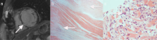
Figure 1
shows (a) the widespread mid-wall LGE in the left ventricle on CMR, (b) lateral wall
fibrosis on the explant specimen (H&E, x20), (c) Giant cell myocarditis in pre-transplant
diagnostic endomyocardial biopsy (H&E, x200) of Patient No: 4 who had CMR and who
subsequently underwent cardiac transplantation.