31
views
0
recommends
+1 Recommend
1 collections
    0
    shares

      International Journal of COPD (submit here)

      This international, peer-reviewed Open Access journal by Dove Medical Press focuses on pathophysiological processes underlying Chronic Obstructive Pulmonary Disease (COPD) interventions, patient focused education, and self-management protocols. Sign up for email alerts here.

      39,063 Monthly downloads/views I 2.893 Impact Factor I 5.2 CiteScore I 1.16 Source Normalized Impact per Paper (SNIP) I 0.804 Scimago Journal & Country Rank (SJR)

      • Record: found
      • Abstract: found
      • Article: found
      Is Open Access

      Alveolar–capillary reserve during exercise in patients with chronic obstructive pulmonary disease

      research-article

      Read this article at

      ScienceOpenPublisherPMC
      Bookmark
          There is no author summary for this article yet. Authors can add summaries to their articles on ScienceOpen to make them more accessible to a non-specialist audience.

          Abstract

          Background

          Factors limiting exercise in patients with COPD are complex. With evidence for accelerated pulmonary vascular aging, destruction of alveolar–capillary bed, and hypoxic pulmonary vasoconstriction, the ability to functionally expand surface area during exercise may become a primary limitation.

          Purpose

          To quantify measures of alveolar–capillary recruitment during exercise and the relationship to exercise capacity in a cohort of COPD patients.

          Methods

          Thirty-two subjects gave consent (53% male, with mean ± standard deviation age 66±9 years, smoking 35±29 pack-years, and Global Initiative for Chronic Obstructive Lung Disease (GOLD) classification of 0–4: 2.3±0.8), filled out the St George’s Respiratory Questionnaire (SGRQ) to measure quality of life, had a complete blood count drawn, and underwent spirometry. The intrabreath (IB) technique for lung diffusing capacity for carbon monoxide (IBDLCO) and pulmonary blood flow (IBQc, at rest) was also performed. Subsequently, they completed a cycle ergometry test to exhaustion with measures of oxygen saturation and expired gases.

          Results

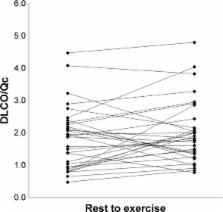
          Baseline average measures were 44±21 for SGRQ score and 58±11 for FEV 1/FVC. Peak oxygen consumption (VO 2) was 11.4±3.1 mL/kg/min (49% predicted). The mean resting IBDLCO was 9.7±5.4 mL/min/mmHg and IBQc was 4.7±0.9 L/min. At the first workload, heart rate (HR) increased to 92±11 bpm, VO 2 was 8.3±1.4 mL/kg/min, and IBDLCO and IBQc increased by 46% and 43%, respectively, compared to resting values ( p,0.01). The IBDLCO/Qc ratio averaged 2.0±1.1 at rest and remained constant during exercise with marked variation across subjects (range: 0.8–4.8). Ventilatory efficiency plateaued at 37±5 during exercise, partial pressure of mix expired CO 2/partial pressure of end tidal CO 2 ratio ranged from 0.63 to 0.67, while a noninvasive index of pulmonary capacitance, O 2 pulse × PetCO 2 (GxCap) rose to 138%. The exercise IBDLCO/Qc ratio was related to O 2 pulse (VO 2/HR, r=0.58, p<0.01), and subjects with the highest exercise IBDLCO/Qc ratio or the greatest rise from rest had the highest peak VO 2 values ( r=0.65 and 0.51, respectively, p<0.05). Of the noninvasive gas exchange measures of pulmonary vascular function, GxCap was most closely associated with DLCO, DLCO/Qc, and VO 2 peak.

          Conclusion

          COPD patients who can expand gas exchange surface area as assessed with DLCO during exercise relative to pulmonary blood flow have a more preserved exercise capacity.

          Most cited references27

          • Record: found
          • Abstract: found
          • Article: not found

          Pulmonary hypertension in chronic lung diseases.

          Chronic obstructive lung disease (COPD) and diffuse parenchymal lung diseases (DPLD), including idiopathic pulmonary fibrosis (IPF) and sarcoidosis, are associated with a high incidence of pulmonary hypertension (PH), which is linked with exercise limitation and a worse prognosis. Patients with combined pulmonary fibrosis and emphysema (CPFE) are particularly prone to the development of PH. Echocardiography and right heart catheterization are the principal modalities for the diagnosis of COPD and DPLD. For discrimination between group 1 PH patients with concomitant respiratory abnormalities and group 3 PH patients (PH caused by lung disease), patients should be transferred to a center with expertise in both PH and lung diseases for comprehensive evaluation. The task force encompassing the authors of this article provided criteria for this discrimination and suggested using the following definitions for group 3 patients, as exemplified for COPD, IPF, and CPFE: COPD/IPF/CPFE without PH (mean pulmonary artery pressure [mPAP] <25 mm Hg); COPD/IPF/CPFE with PH (mPAP ≥25 mm Hg); PH-COPD, PH-IPF, and PH-CPFE); COPD/IPF/CPFE with severe PH (mPAP ≥35 mm Hg or mPAP ≥25 mm Hg with low cardiac index [CI <2.0 l/min/m(2)]; severe PH-COPD, severe PH-IPF, and severe PH-CPFE). The "severe PH group" includes only a minority of chronic lung disease patients who are suspected of having strong general vascular abnormalities (remodeling) accompanying the parenchymal disease and with evidence of an exhausted circulatory reserve rather than an exhausted ventilatory reserve underlying the limitation of exercise capacity. Exertional dyspnea disproportionate to pulmonary function tests, low carbon monoxide diffusion capacity, and rapid decline of arterial oxygenation upon exercise are typical clinical features of this subgroup with poor prognosis. Studies evaluating the effect of pulmonary arterial hypertension drugs currently not approved for group 3 PH patients should focus on this severe PH group, and for the time being, these patients should be transferred to expert centers for individualized patient care.
            Bookmark
            • Record: found
            • Abstract: found
            • Article: not found

            Exercise pathophysiology in patients with primary pulmonary hypertension.

            Patients with primary pulmonary hypertension (PPH) have a pulmonary vasculopathy that leads to exercise intolerance due to dyspnea and fatigue. To better understand the basis of the exercise limitation in patients with PPH, cardiopulmonary exercise testing (CPET) with gas exchange measurements, New York Heart Association (NYHA) symptom class, and resting pulmonary hemodynamics were studied. We retrospectively evaluated 53 PPH patients who had right heart catheterization and cycle ergometer CPET studies to maximum tolerance as part of their clinical workups. No adverse events occurred during CPET. Reductions in peak O(2) uptake (VO(2)), anaerobic threshold, peak O(2) pulse, rate of increase in VO(2), and ventilatory efficiency were consistently found. NYHA class correlated well with the above parameters of aerobic function and ventilatory efficiency but less well with resting pulmonary hemodynamics. Patients with PPH can safely undergo noninvasive cycle ergometer CPET to their maximal tolerance. The CPET abnormalities were consistent and characteristic and correlated well with NYHA class.
              Bookmark
              • Record: found
              • Abstract: not found
              • Article: not found

              Coming together: the ATS/ERS consensus on clinical pulmonary function testing.

              V Brusasco (2005)
                Bookmark

                Author and article information

                Journal
                Int J Chron Obstruct Pulmon Dis
                Int J Chron Obstruct Pulmon Dis
                International Journal of COPD
                International Journal of Chronic Obstructive Pulmonary Disease
                Dove Medical Press
                1176-9106
                1178-2005
                2017
                24 October 2017
                : 12
                : 3115-3122
                Affiliations
                [1 ]Division of Critical Care, Florida Hospital, Orlando, FL
                [2 ]Division of Cardiovascular Diseases, Mayo Clinic, Scottsdale, AZ, USA
                [3 ]Australian School of Advanced Medicine, Faculty of Medicine and Health Sciences, Macquarie University, Sydney, NSW, Australia
                Author notes
                Correspondence: Mehrdad Behnia, Division of Critical Care, Florida Hospital, 601 E Rollins Street, Orlando, FL 32803, USA, Tel +1 706 339 8634, Email doctorbehnia@ 123456gmail.com
                Article
                copd-12-3115
                10.2147/COPD.S142523
                5661445
                29123389
                328d1d86-5a1b-47e6-921d-05763d150337
                © 2017 Behnia et al. This work is published and licensed by Dove Medical Press Limited

                The full terms of this license are available at https://www.dovepress.com/terms.php and incorporate the Creative Commons Attribution – Non Commercial (unported, v3.0) License ( http://creativecommons.org/licenses/by-nc/3.0/). By accessing the work you hereby accept the Terms. Non-commercial uses of the work are permitted without any further permission from Dove Medical Press Limited, provided the work is properly attributed.

                History
                Categories
                Original Research

                Respiratory medicine
                airflow limitation,exercise intolerance,lung gas transfer,dyspnea,copd,diffusion capacity,cardiopulmonary exercise testing

                Comments

                Comment on this article