7
views
0
recommends
+1 Recommend
0 collections
    0
    shares
      • Record: found
      • Abstract: found
      • Article: found
      Is Open Access

      Synapse-specific opioid modulation of thalamo-cortico-striatal circuits

      research-article

      Read this article at

      Bookmark
          There is no author summary for this article yet. Authors can add summaries to their articles on ScienceOpen to make them more accessible to a non-specialist audience.

          Abstract

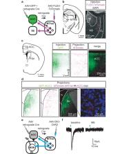
          The medial thalamus (MThal), anterior cingulate cortex (ACC) and striatum play important roles in affective-motivational pain processing and reward learning. Opioids affect both pain and reward through uncharacterized modulation of this circuitry. This study examined opioid actions on glutamate transmission between these brain regions in mouse. Mu-opioid receptor (MOR) agonists potently inhibited MThal inputs without affecting ACC inputs to individual striatal medium spiny neurons (MSNs). MOR activation also inhibited MThal inputs to the pyramidal neurons in the ACC. In contrast, delta-opioid receptor (DOR) agonists disinhibited ACC pyramidal neuron responses to MThal inputs by suppressing local feed-forward GABA signaling from parvalbumin-positive interneurons. As a result, DOR activation in the ACC facilitated poly-synaptic (thalamo-cortico-striatal) excitation of MSNs by MThal inputs. These results suggest that opioid effects on pain and reward may be shaped by the relative selectivity of opioid drugs to the specific circuit components.

          Related collections

          Most cited references46

          • Record: found
          • Abstract: found
          • Article: not found

          Transgenic mice for intersectional targeting of neural sensors and effectors with high specificity and performance.

          An increasingly powerful approach for studying brain circuits relies on targeting genetically encoded sensors and effectors to specific cell types. However, current approaches for this are still limited in functionality and specificity. Here we utilize several intersectional strategies to generate multiple transgenic mouse lines expressing high levels of novel genetic tools with high specificity. We developed driver and double reporter mouse lines and viral vectors using the Cre/Flp and Cre/Dre double recombinase systems and established a new, retargetable genomic locus, TIGRE, which allowed the generation of a large set of Cre/tTA-dependent reporter lines expressing fluorescent proteins, genetically encoded calcium, voltage, or glutamate indicators, and optogenetic effectors, all at substantially higher levels than before. High functionality was shown in example mouse lines for GCaMP6, YCX2.60, VSFP Butterfly 1.2, and Jaws. These novel transgenic lines greatly expand the ability to monitor and manipulate neuronal activities with increased specificity.
            Bookmark
            • Record: found
            • Abstract: found
            • Article: not found

            Functional imaging of brain responses to pain. A review and meta-analysis (2000).

            Brain responses to pain, assessed through positron emission tomography (PET) and functional magnetic resonance imaging (fMRI) are reviewed. Functional activation of brain regions are thought to be reflected by increases in the regional cerebral blood flow (rCBF) in PET studies, and in the blood oxygen level dependent (BOLD) signal in fMRI. rCBF increases to noxious stimuli are almost constantly observed in second somatic (SII) and insular regions, and in the anterior cingulate cortex (ACC), and with slightly less consistency in the contralateral thalamus and the primary somatic area (SI). Activation of the lateral thalamus, SI, SII and insula are thought to be related to the sensory-discriminative aspects of pain processing. SI is activated in roughly half of the studies, and the probability of obtaining SI activation appears related to the total amount of body surface stimulated (spatial summation) and probably also by temporal summation and attention to the stimulus. In a number of studies, the thalamic response was bilateral, probably reflecting generalised arousal in reaction to pain. ACC does not seem to be involved in coding stimulus intensity or location but appears to participate in both the affective and attentional concomitants of pain sensation, as well as in response selection. ACC subdivisions activated by painful stimuli partially overlap those activated in orienting and target detection tasks, but are distinct from those activated in tests involving sustained attention (Stroop, etc.). In addition to ACC, increased blood flow in the posterior parietal and prefrontal cortices is thought to reflect attentional and memory networks activated by noxious stimulation. Less noted but frequent activation concerns motor-related areas such as the striatum, cerebellum and supplementary motor area, as well as regions involved in pain control such as the periaqueductal grey. In patients, chronic spontaneous pain is associated with decreased resting rCBF in contralateral thalamus, which may be reverted by analgesic procedures. Abnormal pain evoked by innocuous stimuli (allodynia) has been associated with amplification of the thalamic, insular and SII responses, concomitant to a paradoxical CBF decrease in ACC. It is argued that imaging studies of allodynia should be encouraged in order to understand central reorganisations leading to abnormal cortical pain processing. A number of brain areas activated by acute pain, particularly the thalamus and anterior cingulate, also show increases in rCBF during analgesic procedures. Taken together, these data suggest that hemodynamic responses to pain reflect simultaneously the sensory, cognitive and affective dimensions of pain, and that the same structure may both respond to pain and participate in pain control. The precise biochemical nature of these mechanisms remains to be investigated.
              Bookmark
              • Record: found
              • Abstract: not found
              • Article: not found

              State-dependent opioid control of pain.

                Bookmark

                Author and article information

                Contributors
                Role: Reviewing Editor
                Role: Senior Editor
                Journal
                eLife
                Elife
                eLife
                eLife
                eLife Sciences Publications, Ltd
                2050-084X
                17 May 2019
                2019
                : 8
                : e45146
                Affiliations
                [1 ]deptVollum Institute Oregon Health & Science University PortlandUnited States
                [2 ]deptDepartment of Anesthesiology Perioperative and Pain Medicine, Stanford Neurosciences Institute Stanford University StanfordUnited States
                [3 ]deptDepartment of Molecular and Cellular Physiology, Stanford Neurosciences Institute Stanford University StanfordUnited States
                [4 ]deptDepartment of Neurosurgery, Stanford Neurosciences Institute Stanford University StanfordUnited States
                [5 ]deptNew York Stem Cell Foundation – Robertson Investigator Stanford University Palo AltoUnited States
                Aix-Marseille University, INSERM, INMED France
                Emory University United States
                Aix-Marseille University, INSERM, INMED France
                University of Colorado Anschutz Medical Campus United States
                UTSA - The University of Texas at San Antonio United States
                Author notes
                [‡]

                Department of Pharmacology, University of Michigan, Ann Arbor, United States.

                [†]

                These authors contributed equally to this work.

                Author information
                https://orcid.org/0000-0003-4799-333X
                https://orcid.org/0000-0002-3532-8319
                Article
                45146
                10.7554/eLife.45146
                6541437
                31099753
                3296247c-d83a-429d-9edc-564a3bb311c7
                © 2019, Birdsong et al

                This article is distributed under the terms of the Creative Commons Attribution License, which permits unrestricted use and redistribution provided that the original author and source are credited.

                History
                : 14 January 2019
                : 15 May 2019
                Funding
                Funded by: FundRef http://dx.doi.org/10.13039/100000065, National Institute of Neurological Disorders and Stroke;
                Award ID: R01NS081071
                Award Recipient :
                Funded by: FundRef http://dx.doi.org/10.13039/100003194, New York Stem Cell Foundation;
                Award Recipient :
                Funded by: FundRef http://dx.doi.org/10.13039/100000026, National Institute on Drug Abuse;
                Award ID: R01DA042779
                Award Recipient :
                Funded by: FundRef http://dx.doi.org/10.13039/100000026, National Institute on Drug Abuse;
                Award ID: R01DA044481
                Award Recipient :
                Funded by: FundRef http://dx.doi.org/10.13039/100000026, National Institute on Drug Abuse;
                Award ID: R01NS106301
                Award Recipient :
                Funded by: FundRef http://dx.doi.org/10.13039/100000065, National Institute of Neurological Disorders and Stroke;
                Award ID: R01NS104944
                Award Recipient :
                Funded by: FundRef http://dx.doi.org/10.13039/100000065, National Institute of Neurological Disorders and Stroke;
                Award ID: U01NS094247
                Award Recipient :
                The funders had no role in study design, data collection and interpretation, or the decision to submit the work for publication.
                Categories
                Research Article
                Neuroscience
                Custom metadata
                By differentially modulating the two major excitatory inputs to the striatum, mu- and delta-opioid receptors regulate the balance between thalamic and cortical inputs to the striatum.

                Life sciences
                opioid,thalamus,striatum,cortex,feed-forward inhibition,opioid receptor,mouse
                Life sciences
                opioid, thalamus, striatum, cortex, feed-forward inhibition, opioid receptor, mouse

                Comments

                Comment on this article