60
views
0
recommends
+1 Recommend
0 collections
    0
    shares
      • Record: found
      • Abstract: found
      • Article: found
      Is Open Access

      Balloon-Occluded Retrograde Transvenous Obliteration for Gastric Variceal Bleeding: Its Feasibility Compared with Transjugular Intrahepatic Portosystemic Shunt

      research-article

      Read this article at

      Bookmark
          There is no author summary for this article yet. Authors can add summaries to their articles on ScienceOpen to make them more accessible to a non-specialist audience.

          Abstract

          Objective

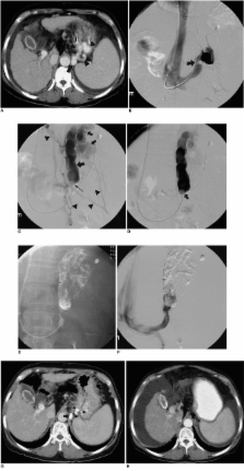
          To assess the feasibility of balloon-occluded retrograde transvenous obliteration (BRTO) in active gastric variceal bleeding, and to compare the findings with those of transjugular intrahepatic portosystemic shunt (TIPS).

          Materials and Methods

          Twenty-one patients with active gastric variceal bleeding due to liver cirrhosis were referred for radiological intervention. In 15 patients, contrast-enhanced CT scans demonstrated gastrorenal shunt, and the remaining six (Group 1) underwent TIPS. Seven of the 15 with gastrorenal shunt (Group 2) were also treated with TIPS, and the other eight (Group 3) underwent BRTO. All patients were followed up for 6 to 21 (mean, 14.4) months. For statistical inter-group comparison of immediate hemostasis, rebleeding and encephalopathy, Fisher's exact test was used. Changes in the Child-Pugh score before and after each procedure in each group were statistically analyzed by means of Wilcoxon's signed rank test.

          Results

          One patient in Group 1 died of sepsis, acute respiratory distress syndrome, and persistent bleeding three days after TIPS, while the remaining 20 survived the procedure with immediate hemostasis. Hepatic encephalopathy developed in four patients (one in Group 1, three in Group 2, and none in Group 3); one, in Group 2, died while in an hepatic coma 19 months after TIPS. Rebleeding occurred in one patient, also in Group 2. Except for transient fever in two Group-3 patients, no procedure-related complication occurred. In terms of immediate hemostasis, rebleeding and encephalopathy, there were no statistically significant differences between the groups ( p > 0.05). In Group 3, the Child-Pugh score showed a significant decrease after the procedure ( p = 0.02).

          Conclusion

          BRTO can effectively control active gastric variceal bleeding, and because of immediate hemostasis, the absence of rebleeding, and improved liver function, is a good alternative to TIPS in patients in whom such bleeding, accompanied by gastrorenal shunt, occurs.

          Related collections

          Most cited references31

          • Record: found
          • Abstract: found
          • Article: not found

          Prevalence, classification and natural history of gastric varices: a long-term follow-up study in 568 portal hypertension patients.

          To determine the prevalence and natural history of gastric varices, we prospectively studied 568 patients (393 bleeders and 175 nonbleeders) with portal hypertension (cirrhosis in 301 patients, noncirrhotic portal fibrosis in 115 patients, extrahepatic portal vein obstruction in 117 patients and hepatic venous outflow obstruction in 35 patients). Primary (present at initial examination) gastric varices were seen in 114 (20%) patients; more were present in bleeders than in non-bleeders (27% vs. 4%, respectively; p < 0.001). Secondary (occurring after obliteration of esophageal varices) gastric varices developed in 33 (9%) patients during follow-up of 24.6 +/- 5.3 mo. Gastric varices (compared with esophageal varices) bled in significantly fewer patients (25% vs. 64%, respectively). Gastric varices had a lower bleeding risk factor than did esophageal varices (2.0 +/- 0.5 vs. 4.3 +/- 0.4, respectively) but bled more severely (4.8 +/- 0.6 vs. 2.9 +/- 0.3 transfusion units per patient, respectively). Once a varix bled, mortality was more likely (45%) in gastric varix patients. Gastric varices were classified as gastroesophageal or isolated gastric varices. Type 1 gastroesophageal varices (lesser curve varices) were the most common (75%). After obliteration of esophageal varices, type 1 gastroesophageal varices disappeared in 59% of patients and persisted in the remainder; bleeding from persistent gastroesophageal varices was more common than it was from gastroesophageal varices that were obliterated (28% vs. 2%, respectively; p < 0.001). Type 2 gastroesophageal varices, which extend to greater curvature, bled often (55%) and were associated with high mortality. Type 1 isolated gastric varices patients had only fundal varices, with a high (78%) incidence of bleeding.(ABSTRACT TRUNCATED AT 250 WORDS)
            Bookmark
            • Record: found
            • Abstract: found
            • Article: not found

            Prediction of variceal hemorrhage by esophageal endoscopy.

            To assess the risk of variceal bleeding, endoscopic findings in 172 patients with esophageal varices were classified based on the "General Rules for Recording Endoscopic Findings on Esophageal Varices" prepared by the Japanese Research Society for Portal Hypertension. Among the factor which enabled an accurate prediction of bleeding were the red wale marking and cherry-red spot of the red color sign category and blue varices of the fundamental color category with large varices. Other factors in the "General Rules" such as the form and location were of minor significance in prediction of the bleeding. Each of these factors can be quantified and an assessment can be made of the likelihood of bleeding. By calculating the total score of these factors, the risk of bleeding in individual patients was accurately estimated.
              Bookmark
              • Record: found
              • Abstract: found
              • Article: not found

              Retrograde transvenous obliteration of gastric varices.

              To evaluate the clinical efficacy, techniques, and complications associated with balloon-occluded retrograde transvenous obliteration of gastric varices. Between December 1994 and November 1997, balloon-occluded retrograde transvenous obliteration was performed on 20 patients with gastric varices in danger of rupture and with gastrorenal shunts; three patients also had hepatic encephalopathy. The sclerosant was injected into the gastric varices during balloon occlusion. The degree of progression of the gastric varices and of collateral veins was classified into five grades, with grade 1 being least progression and grade 5 most progression; collateral veins that had developed were treated with embolization. Follow-up consisted of fiberoptic endoscopy and computed tomography. Technical success was achieved in all patients. Occlusion of collateral veins was essential for the occlusion of gastric varices with a grade greater than grade 2. The clinical symptoms of hepatic encephalopathy in the three patients improved remarkably. Follow-up endoscopy 3 months after the procedure revealed the disappearance of gastric varices in 15 patients and reduced variceal size in five. During the follow-up period, 19 patients had no recurrence of gastric varices; three patients had aggravation of the esophageal varices. Balloon-occluded retrograde transvenous obliteration is a feasible alternative to a transjugular intrahepatic portosystemic shunt for patients with large gastrorenal shunts or hepatic encephalopathy (or both).
                Bookmark

                Author and article information

                Journal
                Korean J Radiol
                KJR
                Korean Journal of Radiology
                The Korean Radiological Society
                1229-6929
                2005-8330
                Apr-Jun 2003
                30 June 2003
                : 4
                : 2
                : 109-116
                Affiliations
                [1 ]Department of Radiology, Seoul City Boramae Hospital, Seoul, Korea.
                [2 ]Department of Radiology, Seoul National University College of Medicine and Institute of Radiation Medicine, Seoul National University Medical Research Center and Clinical Research Institute, Seoul National University Hospital, Seoul, Korea.
                [3 ]Department of Radiology, Cheju National University College of Medicine, Jeju-do, Korea.
                Author notes
                Address reprint requests to: Jin Wook Chung, MD, Department of Radiology, Seoul National University College of Medicine, 28 Yongon-dong, Chongno-gu, Seoul 110-744, Korea. Telephone: (822) 760-2584, Fax: (822) 743-6385, chungjw@ 123456radcom.snu.ac.kr
                Article
                10.3348/kjr.2003.4.2.109
                2698068
                12845306
                32f33106-86f2-4b63-992d-bbbefc6eaac6
                Copyright © 2003 The Korean Radiological Society

                This is an Open Access article distributed under the terms of the Creative Commons Attribution Non-Commercial License ( http://creativecommons.org/licenses/by-nc/3.0) which permits unrestricted non-commercial use, distribution, and reproduction in any medium, provided the original work is properly cited.

                History
                : 14 August 2002
                : 10 March 2003
                Categories
                Original Article

                Radiology & Imaging
                hypertension, portal,stomach, varices,veins, therapeutic blockade,interventional procedures, technology,stomach, bleeding

                Comments

                Comment on this article