7
views
0
recommends
+1 Recommend
0 collections
    0
    shares
      • Record: found
      • Abstract: found
      • Article: found
      Is Open Access

      Changes in choroidal blood flow velocity in patients diagnosed with central serous chorioretinopathy during follow-up for pachychoroid pigment epitheliopathy

      case-report

      Read this article at

      Bookmark
          There is no author summary for this article yet. Authors can add summaries to their articles on ScienceOpen to make them more accessible to a non-specialist audience.

          Abstract

          Purpose

          To evaluate chronological changes in choroidal blood flow velocity in two patients with pachychoroid pigment epitheliopathy (PPE) and central serous chorioretinopathy (CSC) in the same eye.

          Observations

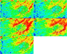
          Two males aged 36 and 43 years old with PPE were diagnosed with CSC in the same eyes during follow-up. Using laser speckle flowgraphy, the macular mean blur rate (MBR), an index of relative blood flow velocity, was sequentially evaluated in the affected and unaffected eyes. In the affected eye, the macular MBR values at the onset of PPE and CSC were higher, at 25% and 33% in Case 1 and 21% and 51% in Case 2, respectively, than those on PPE regression; but the same trends were not observed in their fellow eyes. The increases in MBR changing rates were 1.3 and 2.5 times higher in Cases 1 and 2, respectively, at the onset of CSC than those at the onset of PPE.

          Conclusion and importance

          In the affected eyes, the rates of MBR change increased at the alternate onsets of PPE and CSC. The increased MBR changing rates were 1.3–2.5 times higher at the onset of CSC than those at the onset of PPE. Our data suggest that choroidal hyperperfusion is involved in the pathogenesis of both diseases and that its severity may differ between CSC and PPE. These results may support the hypothesis that PPE and CSC clinically overlap and have a common pathogenic background.

          Related collections

          Most cited references24

          • Record: found
          • Abstract: found
          • Article: not found

          Pachychoroid pigment epitheliopathy.

          To report nine cases of pachychoroid pigment epitheliopathy. An observational case series of nine patients who underwent comprehensive ophthalmic examination, fundus photography, fundus autofluorescence, spectral-domain optical coherence tomography, and enhanced depth imaging optical coherence tomography. Eighteen eyes of 9 patients, aged 27 years to 89 years, were diagnosed with pachychoroid pigment epitheliopathy based on the characteristic funduscopic appearance of reduced fundus tessellation with overlying retinal pigment epithelial changes in one or both eyes, fundus autofluorescence abnormalities, and increased subfoveal choroidal thickness confirmed by enhanced depth imaging optical coherence tomography (mean, 460.2 μm). The five older patients had been previously diagnosed with age-related macular degeneration, while the four younger subjects were referred for possible inflammatory chorioretinitis, pattern dystrophy, or nonspecific drusen. No subjects had a history of or subsequently developed subretinal fluid. Pachychoroid pigment epitheliopathy falls within a spectrum of diseases associated with choroidal thickening that includes central serous chorioretinopathy and polypoidal choroidal vasculopathy, and it should be suspected in eyes with a characteristic fundus appearance related to choroidal thickening and associated retinal pigment epithelial abnormalities but no history of subretinal fluid. Enhanced depth imaging optical coherence tomography confirming an abnormally thick choroid and characteristic retinal pigment epithelial changes on fundus autofluorescence support the diagnosis. Because these patients are frequently misdiagnosed, the recognition of pachychoroid pigment epitheliopathy may avoid unnecessary diagnostic testing and interventions.
            Bookmark
            • Record: found
            • Abstract: found
            • Article: not found

            Pachychoroid disease

            Pachychoroid is a relatively novel concept describing a phenotype characterized by attenuation of the choriocapillaris overlying dilated choroidal veins, and associated with progressive retinal pigment epithelium dysfunction and neovascularization. The emphasis in defining pachychoroid-related disorders has shifted away from simply an abnormally thick choroid (pachychoroid) toward a detailed morphological definition of a pathologic state (pachychoroid disease) with functional implications, which will be discussed in this review. Several clinical manifestations have been described to reside within the pachychoroid disease spectrum, including central serous chorioretinopathy, pachychoroid pigment epitheliopathy, pachychoroid neovasculopathy, polypoidal choroidal vasculopathy/aneurysmal type 1 neovascularization, focal choroidal excavation, peripapillary pachychoroid syndrome. These conditions all exhibit the characteristic choroidal alterations and are believed to represent different manifestations of a common pathogenic process. This review is based on both the current literature and the clinical experience of our individual authors, with an emphasis on the clinical and imaging features, management considerations, as well as current understanding of pathogenesis of these disorders within the context of the recent findings related to pachychoroid disease.
              Bookmark
              • Record: found
              • Abstract: found
              • Article: not found

              EN FACE IMAGING OF PACHYCHOROID SPECTRUM DISORDERS WITH SWEPT-SOURCE OPTICAL COHERENCE TOMOGRAPHY.

              To correlate clinical manifestations with choroidal morphology in pachychoroid disorders, including central serous chorioretinopathy, pachychoroid pigment epitheliopathy, pachychoroid neovasculopathy, and polypoidal choroidal vasculopathy, using en face swept-source optical coherence tomography (OCT).
                Bookmark

                Author and article information

                Contributors
                Journal
                Am J Ophthalmol Case Rep
                Am J Ophthalmol Case Rep
                American Journal of Ophthalmology Case Reports
                Elsevier
                2451-9936
                2451-9936
                05 March 2020
                June 2020
                05 March 2020
                : 18
                : 100651
                Affiliations
                [a ]Department of Ophthalmology, Faculty of Medicine and Graduate School of Medicine, Hokkaido University, Sapporo, Japan
                [b ]Kaimeido Eye and Dental Clinic, Sapporo, Japan
                Author notes
                []Corresponding author. Department of Ophthalmology, Faculty of Medicine and Graduate School of Medicine, Hokkaido University, Nishi 7, Kita 15, Kita-ku, Sapporo, 060-8638, Japan. wsaito@ 123456med.hokudai.ac.jp
                Article
                S2451-9936(20)30032-3 100651
                10.1016/j.ajoc.2020.100651
                7090241
                343a479b-685f-43f1-b208-f1957b58b0a0
                © 2020 The Authors

                This is an open access article under the CC BY-NC-ND license (http://creativecommons.org/licenses/by-nc-nd/4.0/).

                History
                : 6 August 2019
                : 23 December 2019
                : 3 March 2020
                Categories
                Case Report

                central serous chorioretinopathy,choroidal blood flow velocity,laser speckle flowgraphy,mean blur rate,pachychoroid pigment epitheliopathy

                Comments

                Comment on this article