32
views
0
recommends
+1 Recommend
0 collections
    0
    shares
      • Record: found
      • Abstract: found
      • Article: found
      Is Open Access

      Proteomic Analysis of Nasal Epithelial Cells from Cystic Fibrosis Patients

      research-article

      Read this article at

      Bookmark
          There is no author summary for this article yet. Authors can add summaries to their articles on ScienceOpen to make them more accessible to a non-specialist audience.

          Abstract

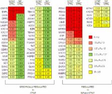
          The pathophysiology of cystic fibrosis (CF) lung disease remains incompletely understood. New explanations for the pathogenesis of CF lung disease may be discovered by studying the patterns of protein expression in cultured human nasal epithelial cells (HNEC). To that aim, we compared the level of protein expressions in primary cultures of HNEC from nasal polyps secondary to CF (CFNP, n = 4), primary nasal polyps (NP, n = 8) and control mucosa (CTRL, n = 4) using isobaric tag for relative and absolute quantification (iTRAQ) labeling coupled with liquid chromatography (LC)-MS-MS. The analysis of the data revealed 42 deregulated protein expressions in CFNP compared to NP and CTRL, suggesting that these alterations are related to CF. Overall, AmiGo analysis highlighted six major pathways important for cell functions that seem to be impaired: metabolism, G protein process, inflammation and oxidative stress response, protein folding, proteolysis and structural proteins. Among them, glucose and fatty acid metabolic pathways could be impaired in CF with nine deregulated proteins. Our proteomic study provides a reproducible set of differentially expressed proteins in airway epithelial cells from CF patients and reveals many novel deregulated proteins that could lead to further studies aiming to clarify the involvement of such proteins in CF pathophysiology.

          Related collections

          Most cited references33

          • Record: found
          • Abstract: found
          • Article: not found

          Identification of the cystic fibrosis gene: chromosome walking and jumping.

          An understanding of the basic defect in the inherited disorder cystic fibrosis requires cloning of the cystic fibrosis gene and definition of its protein product. In the absence of direct functional information, chromosomal map position is a guide for locating the gene. Chromosome walking and jumping and complementary DNA hybridization were used to isolate DNA sequences, encompassing more than 500,000 base pairs, from the cystic fibrosis region on the long arm of human chromosome 7. Several transcribed sequences and conserved segments were identified in this cloned region. One of these corresponds to the cystic fibrosis gene and spans approximately 250,000 base pairs of genomic DNA.
            Bookmark
            • Record: found
            • Abstract: found
            • Article: not found

            Coomassie staining as loading control in Western blot analysis.

            In Western blotting, immunodetection of housekeeping proteins is routinely performed to detect differences in electrophoresis loading. The present work describes a much faster and simpler protein staining method, which is compatible with ordinary blocking conditions. In addition, the method can be used after immunodetection with superior linearity compared to ordinary staining methods. After immunoblotting and staining, protein bands can be further identified using peptide mass fingerprinting.
              Bookmark
              • Record: found
              • Abstract: found
              • Article: not found

              ER chaperone functions during normal and stress conditions.

              Nearly all resident proteins of the organelles along the secretory pathway, as well as proteins that are expressed at the cell surface or secreted from the cell, are first co-translationally translocated into the lumen of the endoplasmic reticulum (ER) as unfolded polypeptide chains. Immediately after entering the ER, they are often modified with N-linked glycans, are folded into the appropriate secondary and tertiary structures, which are stabilized by disulfide bonds, and finally in many cases are assembled into multimeric complexes. These processes are aided and monitored by ER chaperones and folding enzymes. When cells experience conditions that alter the ER environment, protein folding can be dramatically affected and can lead to the accumulation of unfolded proteins in this organelle. This in turn activates a signaling response, which is shared among all eukaryotic organisms, termed the unfolded protein response (UPR). The hallmark of this response is the coordinate transcriptional up-regulation of ER chaperones and folding enzymes. A major role for the increased levels of chaperones and folding enzymes during conditions of ER stress is to provide the same functions they carry out during normal physiological conditions. This includes preventing unfolded and incompletely folded proteins from aggregating and promoting the proper folding and assembly of proteins in the ER. During conditions of ER stress, many proteins are unable to fold properly and the requirements for chaperones are therefore increased. However, more recently it has become clear that some ER chaperones are also involved in signaling the ER stress response, targeting misfolded proteins for degradation and perhaps even shutting down the UPR when the stress subsides. In addition, during some normal physiological conditions, like plasma cell differentiation where there is an increased demand in the secretory capacity of B cells, the levels of various ER chaperones are also up-regulated via at least part of the UPR pathway. In order to discuss these various functions of ER chaperones, we will begin with the roles of ER chaperones and folding enzymes during normal physiological conditions and then discuss their roles during ER stress.
                Bookmark

                Author and article information

                Contributors
                Role: Editor
                Journal
                PLoS One
                PLoS ONE
                plos
                plosone
                PLoS ONE
                Public Library of Science (San Francisco, USA )
                1932-6203
                2014
                30 September 2014
                : 9
                : 9
                : e108671
                Affiliations
                [1 ]Service de Génétique et Embryologie Médicales, Unité Mixte de Recherche_Scientifique 933, Institut National de la Santé et de la Recherche Médicale, Université Pierre et Marie Curie – Paris 6, and Assistance Publique – Hôpitaux de Paris, Hôpital Armand Trousseau, Paris, France
                [2 ]Unité_1151, Institut National de la Santé et de la Recherche Médicale, Université Paris Descartes, Paris, France
                [3 ]Plateau Proteome Necker, Structure Fédérative de Recherche de Necker, Université Paris Descartes, Paris, France
                [4 ]Unité Mixte de Recherche_Scientifique 855, Institut National de la Santé et de la Recherche Médicale, Université Paris 12, Faculté de Médecine, Créteil, France
                [5 ]Service d’Otorhinolaryngologie et de chirurgie cervico-faciale, Assistance Publique – Hôpitaux de Paris, Hôpital inter-communal et Groupe Hospitalier Henri Mondor-Albert Chenevier, Créteil, France
                Shantou University Medical College, China
                Author notes

                Competing Interests: The authors have declared that no competing interests exist.

                Conceived and designed the experiments: AE EE LJ ICG. Performed the experiments: LJ ICG CC. Analyzed the data: ICG LJ. Contributed reagents/materials/analysis tools: AC JFP VPE PZ. Contributed to the writing of the manuscript: LJ ICG AE EE SA AC.

                Article
                PONE-D-14-22452
                10.1371/journal.pone.0108671
                4182543
                25268127
                35389ccd-a6bd-4b3b-a7d7-b6f798e400bd
                Copyright @ 2014

                This is an open-access article distributed under the terms of the Creative Commons Attribution License, which permits unrestricted use, distribution, and reproduction in any medium, provided the original author and source are credited.

                History
                : 5 June 2014
                : 24 August 2014
                Page count
                Pages: 12
                Funding
                Funding was provided by Agence Nationale pour la Recherche -05-MRAR-02201; European Community: Life Sciences and Health Grant (LSHG-CT-2005-512044) NEUPROCF; French Foundation for Cystic Fibrosis: “Vaincre la Mucoviscidose”; Legs Poix from Chancellerie des Universités. The funders had no role in study design, data collection and analysis, decision to publish, or preparation of the manuscript.
                Categories
                Research Article
                Biology and Life Sciences
                Anatomy
                Biological Tissue
                Epithelium
                Biochemistry
                Proteomics
                Developmental Biology
                Fibrosis
                Cystic Fibrosis
                Physical Sciences
                Chemistry
                Analytical Chemistry
                Mass Spectrometry
                Custom metadata
                The authors confirm that all data underlying the findings are fully available without restriction. All relevant data are within the paper and its Supporting Information files.

                Uncategorized
                Uncategorized

                Comments

                Comment on this article